Location: Home >> Detail
Pharm Front. 2019;1:e190007. https://doi.org/10.20900/pf20190007
 ,
Alexandra Francian 1,
Ameneh Arabi 1,
Troy Olsson 2,
Kristine Mann 2,
Holly A. Martinson 1
,
Alexandra Francian 1,
Ameneh Arabi 1,
Troy Olsson 2,
Kristine Mann 2,
Holly A. Martinson 1
1 WWAMI School of Medical Education, University of Alaska Anchorage, 3211 Providence Drive, Anchorage, AK 99508, USA
2 Department of Biological Sciences, University of Alaska Anchorage, Anchorage, AK 99508, USA
* Correspondence: Max Kullberg, Tel.: +1-907-786-7708.
This article belongs to the Virtual Special Issue "Controlled and Targeted Release of Natural Drugs"
In the tumor microenvironment, cytokines, growth factors, and oncogenes mediate constitutive activation of the signal transducer and activator of transcription 3 (STAT3) signaling pathway in both cancer cells and infiltrating immune cells. STAT3 activation in cancer cells drives tumorigenic changes that allow for increased survival, proliferation, and resistance to apoptosis. The modulation of immune cells is more complicated and conflicting. STAT3 signaling drives the myeloid cell phenotype towards an immune suppressive state, which mediates T cell inhibition. On the other hand, STAT3 signaling in T cells leads to proliferation and T cell activity required for an anti-tumor response. Targeted delivery of STAT3 inhibitors to cancer cells and myeloid cells could therefore improve therapeutic outcomes. Many compounds that inhibit the STAT3 pathways for cancer treatment include peptide drugs, small molecule inhibitors, and natural compounds. However, natural compounds that inhibit STAT3 are often hydrophobic, which reduces their bioavailability and leads to unfavorable pharmacokinetics. This review focuses specifically on liposome-encapsulated natural STAT3 inhibitors and their ability to target cancer cells and myeloid cells to reduce tumor growth and decrease STAT3-mediated immune suppression. Many of these liposome formulations have led to profound tumor reduction and examples of combination formulations have been shown to eliminate tumors through immune modulation.
The signal transducer and activator of transcription 3 (STAT3) signaling pathway is often constitutively activated in tumor cells of breast, colorectal, prostate, glioblastoma and other types of cancers [1]. In the tumor microenvironment, cytokines and growth factors that are commonly upregulated such as IL-6, IL-10, PGE2, GM-CSF, VEGF, and EGF and HGF family members activate STAT3 by phosphorylation [2,3]. Oncogenic proteins, Src and Ras, and carcinogens such as tobacco, UVB and LPS have all been shown to activate STAT3 [4]. STAT3 constitutive activity in tumors drives inflammation, apoptosis, cell survival, proliferation, cellular transformation, angiogenesis, metastasis and epithelial to mesenchymal transition (EMT) in cancer progression [5,6]. STAT3 has therefore emerged as a key therapeutic target in the prevention and treatment of cancer [4,7,8].
In the tumor microenvironment, STAT3 activation can result in conflicting immune modulation between myeloid cells and T cells [2,9]. With respect to myeloid cells, STAT3 activation induces tolerogenic activities in tumor-associated myeloid cells such as myeloid derived suppressor cells (MDSCs) and M2 macrophages [10]. STAT3 impairs myeloid cell differentiation, antigen presentation, and promotes immunosuppressive and proangiogenic potential through the secretion of cytokines (IL-10, TGF-B, VEGF) and modulation of metabolic processes (Reactive Oxygen Species, Arginase 1, IDO-1) all which can interfere with T cell activity [11,12]. However, with respect to T cells, STAT3 activation corresponds with proliferation and is a necessary component of a healthy T cell immune response [13,14]. Because of the necessity of STAT3 signaling for an anti-tumor T cell immune response, the most effective delivery of STAT3 inhibitors would be through a mechanism that targets tumor cells and myeloid cells, without delivering to T cells. Nanoparticle delivery is one possibility for increasing specificity of STAT3 inhibition to myeloid and tumor cells.
Because of the profound effect STAT3 activation has on tumor growth, there has been a focus for the last 20 years on creating STAT3 inhibitors for the treatment of cancer [7]. Inhibitor drugs fall into four groups; peptide, small molecule, natural compounds, and tyrosine kinase inhibitors [15]. Many of these STAT3 inhibitors have high cytotoxic IC50 values and in particular the natural compounds are restricted by their poor solubility in water and limited bioavailability, which has prohibited further development. Thus, targeted delivery with nanoparticles has been explored as a possible delivery mechanism to overcome the low bioavailability and to avoid or eliminate adverse effects [16].
Many nanoparticle formulations exist for encapsulation of drugs, including micelles, gold nanoparticles, silicon, liposomes and PGLA formulations. This review will focus on liposomes, which are the most commonly utilized nanoparticle and the only nanoparticle that has FDA approved formulations [17]. Liposomes can be formed from phospholipids that are found naturally in the body and therefore can be metabolized with limited toxicity. Liposomes contain a lipid bilayer, allowing for the encapsulation of hydrophobic compounds, such as many of the natural compound STAT3 inhibitors [18]. In addition, the aqueous lumen of the liposomes allows for the encapsulation of immune activating compounds such as TLR agonists, CpG, poly (I:C) and R848 [19]. Finally, liposomes can be easily modified with targeting ligands, sugars and other moieties to allow for specific uptake into tumor cells or myeloid cells [20]. Therefore, liposome delivery of STAT3 inhibitors directly to the tumor site could be an effective way to target both tumor cells and suppressive myeloid cells, while reducing STAT3-specific toxicity in normal tissues. In this review we will provide an analysis of studies that encapsulated STAT3 inhibitors into liposomes for the purpose of cancer treatment.
Overview of Liposomal STAT3 InhibitorsMultiple therapeutic approaches have been tested to target and inhibit STAT3 signaling, including peptide and small molecule inhibitors that have been formulated to target the SH2 domain of STAT3, thereby inhibiting STAT3 activation [21]. Many of these peptide and small molecule drugs are in various stages of clinical trials, but none have been incorporated into liposomal formulations for therapeutic delivery, and therefore will not be discussed here. The majority of liposomal formulations available for targeting STAT3 are based on natural compounds. For comparison, these natural compounds, the structure of each, the cancers treated, and the significant findings are summarized in Table 1 below.
For each of the studies reviewed, the formulation of phospholipids, the cholesterol content and the percentage of polyethylene glycol conjugated lipids differed. These differences can significantly impact treatment efficacy by affecting pharmacokinetics of drug release and uptake profiles into phagocytic cells [22]. However, given the limited number of studies on liposomal delivery for each natural STAT3 inhibitor and the various cancer models that rarely match between studies, it was not possible to evaluate the effect of liposome compositions on drug efficacy. As more studies emerge on liposomal delivery of STAT3 inhibitors, hopefully the effect of lipid composition on cancer treatment can be adequately addressed.
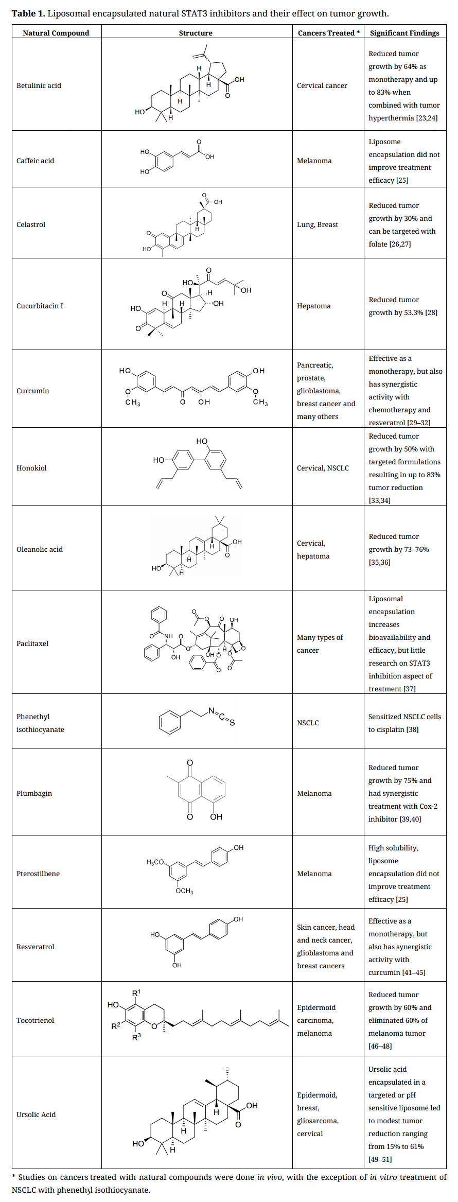 Table 1. Liposomal encapsulated natural STAT3 inhibitors and their effect on tumor growth.
The liposomal formulations of these natural compounds are reviewed below, providing a summary of their activity for STAT3, liposomal encapsulation efficiency, and a discussion of the treatment strategy and effectiveness for the various types of cancer.
Betulinic acidBetulinic acid is a pentacyclic triterpene isolated from many fruits, vegetables, plants, and the bark of birch, sycamore, and eucalyptus trees. Inhibition of STAT3 by betulinic acid occurs by the blocking of nuclear translocation [52,53]. Given its poor water solubility (20 mg/L) and proven efficacy against cancer, betulinic acid is an appropriate candidate for encapsulation in liposomes. Betulinic acid was encapsulated within pegylated liposomes, with an encapsulation efficiency of up to 95%. Mice bearing U14 cervical cancer tumors were treated intratumorally with betulinic acid liposomes, which resulted in a significant tumor inhibition rate of 64%, compared to non-encapsulated betulinic acid (31%). There was no evidence of toxicity as measured by weight loss and behavior [23]. Another study by the same group examined the encapsulation of betulinic acid into gold shell coated liposomes for the purpose of drug delivery combined with photothermal therapy. When used to deliver betulinic acid and heat tumors through near infrared irradiation, liposomes reduced tumor growth by 83% [24]. Although there are limited studies on betulinic acid in liposomal formulations for the treatment of cancer, these results show the possibility of enhancing cancer treatment with liposomal encapsulation and direct administration to the tumor.
Caffeic acidCaffeic acid is a polyphenolic cinnamic acid derivative that is found in the majority of plants, particularly in Eucalyptus globulus, Dipsacus asperoides, and plants of the genus Phellinus. Caffeic acid and its synthetic derivative CADPE (3-(3,4-Dihydroxy-phenyl)-acrylic acid 2-(3,4-dihydroxy-phenyl)-ethyl ester) are both active STAT3 inhibitors. In vitro, caffeic acid has been shown to directly inhibit NF-κB and phosphorylation of STAT3 in response to IL-6 [54]. Both caffeic acid and CAPDE have been shown to reduce tumor growth and angiogenesis by inhibiting the activity of STAT3, HIF-1α, and VEGF in a mouse xenograft model [55]. Caffeic acid has a water solubility of 650 mg/L, representing one of the less hydrophobic compounds of the natural STAT3 inhibitors.
Perhaps because of its relatively high solubility, only one study investigated liposomal targeting of caffeic acid in cancer [25]. Liposome encapsulation efficiency was 2%, which is much lower than many of the drugs reviewed here, most likely because the drug does not partition as readily into the hydrophobic bilayer. Researchers found that the liposome formulation did not result in decreased growth of B16-F10 melanoma cells compared to control liposomes without caffeic acid.
CelastrolCelastrol is a triterpenoid compound derived from the medicinal herb, Tripterygium wilfordii [26,56]. It has been studied extensively for its anti-inflammatory and antioxidant activities and has gained interest recently due to its potential anti-cancer effects in numerous cancer cell lines, including breast, prostate, lung, glioma, myeloma, leukemia, melanoma, and pancreatic cancer [26,27,56]. Celastrol exhibits anticancer activity through inhibition of a variety of biological processes including NF-κB activation, constitutive and IL-6 dependent STAT3 signaling, and VEGF receptor expression, among others [27,57–60]. Celastrol has also been documented as an adjuvant therapy to doxorubicin and paclitaxel chemotherapeutic agents [61]. Clinical application has been limited due to its low aqueous solubility and permeability, poor bioavailability, and systemic toxicity, which necessitates the use of toxic solvents for administration [62,63].
Several studies to date have shown that liposomal formulations of celastrol lower toxicity while enhancing antitumor efficacy of treatments [26,27,56]. Celastrol has been encapsulated in several types of liposome formulations, including pegylated [56,58], cholesterol [26], folate-targeted [57], and microemulsions [64], as well as encapsulated with other drugs (irinotecan, sodium tanshinone IIA sulfonate, and axitinib) [57,58,64]. The encapsulation efficiencies were all high (ranging from 71.67% up to 99.9%), due to the hydrophobic nature of celastrol which allows it to be contained within the lipid bilayer, or oil microemulsion [64].
Improved cytotoxicity, anti-proliferative and anti-apoptotic activity from liposomal celastrol was seen in vitro in several different cell lines, including prostate cancer, breast cancer, glioma, neuroblastoma, and squamous carcinoma [27,56–58,64]. In vivo liposomal delivery of celastrol using several types of xenograft mouse models reported promising results in regard to tumor inhibition. The most impressive tumor inhibition rates were reported in mouse models of Lewis lung cancer [26] and SHG-44 human glioma [27] (34.9% and 32.8%, respectively). These studies demonstrate that celastrol can be effectively encapsulated into targeted and non-targeted liposomes and act synergistically with other drugs to reduce tumor growth in several different in vitro and in vivo cancer models.
CucurbitacinCucurbitacin is a triterpene found in many plants, including cucumbers and pumpkins, with letters A through T denoting 18 variations of the compound. Cucurbitacin has been shown to inhibit proliferation and induce apoptosis in many in vitro and in vivo cancer models as reviewed elsewhere [65]. The anticancer and immunomodulatory effects of cucurbitacin have been linked to its ability to downregulate STAT3 [66,67]. Cucurbitacin-I blocked IL-6 induced STAT3 activation at a concentration of 0.5 µmol/L [68]. Interestingly, cucurbitacin-I also inhibited the dephosphorylation of STAT1, showing that the compound could potentiate STAT1 activation, which is important for immune stimulation. Cucurbitacin is hydrophobic, with a poor water solubility of 25 mg/L and systemic toxicity at therapeutically relevant levels, which limit its effectiveness.
Cucurbitacin has been encapsulated in liposomes with 85% encapsulation efficiency. Lipid nanoparticle encapsulated cucurbitacin-B had uptake that was 1.94 times higher than free cucurbitacin-B in liver cells, suggesting that encapsulating cucurbitacin could improve biodistribution of the drug for anticancer treatment [28]. Cucurbitacin has also been encapsulated in lipid nanoparticles to treat h22 murine hepatoma tumors with intravenous administration resulting in 53.4% reduction in tumor growth, compared to 31.5% with non-encapsulated cucurbitacin [28]. Polymeric micelles have also been used to encapsulate cucurbitacin to treat B16-F10 tumors, which resulted in significant tumor regression [69]. Although there have been limited studies on cucurbitacin in liposome formulations, given its potent inhibition of STAT3 and potentiation of STAT1 signaling, it has potential for immunomodulation in cancer.
CurcuminThe polyphenol, curcumin, is the principle active component of turmeric and has long been used as an herbal and dietary supplement. It has garnered interest in biomedical research due to its anti-infectious, anti-inflammatory, antioxidant, cardio-protective, chemo-preventative, and anticancer properties as well as its low toxicity. Curcumin acts through the modulation of numerous cell signaling molecules and pathways, most notably NF-κB and STAT3, and others such as MAPK, LOX, IL-1β, IL-6, Akt, bcl-2, COX-2. The STAT3 inhibiting properties of curcumin have been well established, with reduction in tumor cell growth that is reversible with the forced overexpression of STAT3 [70–76]. It has also been shown to strongly inhibit the transcription factor NF-κB and related pathways [32,71,77,78]. Curcumin has a unique immunomodulatory function and when delivered in a mouse model of metastatic breast cancer, was shown to repolarize macrophages towards an M1 phenotype, leading to reduced tumor growth [79]. Given the use of curcumin in cancer models and clinical trials, coupled with its poor aqueous solubility (<0.125 mg/L), low bioavailability, light sensitivity, and rapid metabolism, curcumin is an ideal candidate for liposomal encapsulation [71].
Of all the natural STAT3 inhibitors investigated, curcumin in liposomal formulations has been the most extensively studied for the treatment of cancer, with over 140 publications and extensive reviews elsewhere [71]. Briefly, curcumin liposomes have been ligand-modified (vitamin A, folate, hyaluronic acid, epithelial growth factor, integrins, legumain) to direct liposomes to specific receptors [72,74,80–83] or encapsulated with other therapeutic agents (paclitaxel, cisplatin, resveratrol, C6 ceramide, doxorubicin, gemcitabine) to sensitize cancer cells [31,75,84–88]. It is well-tolerated at high doses and has been encapsulated in multiple liposomal formulations with efficiencies ranging from approximately 50–99%. Liposomal curcumin has been used in clinical trials and against several different cancer cell lines, such as lung, glioblastoma, cervical, prostate, breast, osteosarcoma, liver, and head and neck cancers [71,89].
Several studies have determined that curcumin acts through inhibition of STAT3 phosphorylation or related pathways. In a study by Mondal et al., curcumin was encapsulated in a liposome targeted to the tumor vasculature through integrin receptors, with encapsulation efficiencies ranging from 85% to 90% [74]. These liposomes showed significant anticancer activity in endothelial and melanoma cancer cell lines. In a syngeneic melanoma mouse model, tumor growth was inhibited by intravenous treatment with the targeted liposomes. Immunohistochemistry analysis revealed that tumor growth inhibition was mediated by blocking VEGF-induced STAT3 phosphorylation in the tumor endothelium [74]. In a study by Amano et al., curcumin was encapsulated in liposomes and used to determine the effects on macrophages. Intraperitoneal treatment of mice resulted in high uptake by macrophages and significant reduction of IL-6 production [76]. In a study by Zhang et al., hydrazinocurcumin (HC), a synthetic analog of curcumin, was targeted to macrophages with legumain. Previously, HC was determined to be an effective inhibitor of STAT3 phosphorylation. Targeted liposomes were successfully able to suppress STAT3 activity in tumor associated macrophages (TAMs) and re-polarize them to an anti-tumor M1-like phenotype. In vivo, treatment resulted in suppression of tumor growth, metastasis, and angiogenesis [72].
Curcumin has been encapsulated in liposomes with several other phytochemicals, chemotherapies, and compounds. A synthetic analog of curcumin, CDF, was encapsulated in a liposome and used to treat cisplatin-resistant head and neck cancer. In a xenograft mouse model, liposome and cisplatin treatment resulted in significant tumor reduction and growth inhibition, as well as reduction of the expression of CD44 (cancer stem cell marker) [85]. Several studies have shown that curcumin may benefit from addition of a chemotherapeutic agent. When used to treat a mouse model of cervical cancer, liposomal curcumin alone did not have an antitumor effect. However, when combined with paclitaxel, there was a synergistic effect that the authors hypothesized was a result of curcumin downregulating anti-apoptotic survival signals, such as NF-κB [32]. In a study by Sesarman et al., curcumin was encapsulated together with DOX in a pegylated liposome formulation with an encapsulation efficiency of 87.48%. These liposomes were used to treat murine colon carcinoma cells. Treatment resulted in increased anticancer activity compared to single agents, and slightly increased compared to free curcumin and DOX combined [88]. At least three in vivo studies have examined the possibility of co-encapsulating curcumin and resveratrol in a liposomal formulation for cancer treatment. Curcumin and resveratrol liposomes were used to treat male prostate-specific PTEN knockout mice, a model for human prostate cancer. In vitro, the liposome treatment resulted in inhibition of cell growth and increased apoptosis. In vivo, treatment with the multidrug liposomes significantly reduced the occurrence of prostate adenocarcinomas [31]. Alone, liposomal curcumin or resveratrol resulted in approximately 50% reduction in growth, but when co-encapsulated, they reduced tumor growth by close to 75%. When curcumin, resveratrol and epicatechin gallate were used to treat glioblastoma, the tumors were eliminated due to a repolarization of the tumor associated macrophages [30]. Using the same formulation, the authors treated tumors derived from HNSCC cells, and again showed that increased STAT1 signaling and inhibition of STAT3 signaling polarized macrophages to an M1 phenotype and corresponded with an 86% decrease in tumor growth [90].
In a phase 1 study, 32 patients with metastatic cancer were treated with liposomal curcumin (Lipocurc™). Although there was no observed tumor reduction, liposomal curcumin was generally well tolerated and no dose-limiting toxicity was observed in 26 patients at doses between 100 and 300 mg/m2 over 8 h [89].
These studies suggest liposome encapsulation provides an effective delivery system to enhance and expand the therapeutic potential of curcumin by improving pharmacokinetics and combination therapies.
HonokiolHonokiol is a polyphenol isolated from the bark and leaves of trees of the genus Magnolia. Used in traditional Chinese and Japanese medicine, Honokiol is known to possess anti-thrombocytic, antioxidant, antibacterial, anti-inflammatory, and antiproliferative effects. Honokiol has been used to treat many types of cancers, with its anticancer activity attributed at least partially to its potent inhibition of STAT3 phosphorylation [91,92]. Honokiol is hydrophobic and has a water solubility of 53 mg/mL.
Honokiol has been extensively studied in liposomal formulations for the treatment of glioblastoma, colon cancer, breast cancer, ovarian cancer and non-small cell lung cancer [33,34,93,94]. Many of these studies come from the same lab, which has combined liposomal honokiol with chemotherapeutics such as cisplatin and epirubicin. When administered intraperitoneally in combination with cisplatin, liposomal honokiol increased sensitivity to treatment, resulting in 88% inhibition of tumor growth in a cisplatin-resistant ovarian cancer model [34]. There was no evidence of toxicity as measured by weight loss, behavior, and H&E histological staining of all major organs [34]. Another combinational therapy, encapsulating both epirubicin and honokiol, was targeted to somatostatin receptors in non-small cell lung cancer using the ligand octreotide [33]. This liposome formulation had an encapsulation efficiency of 96%. Treatment resulted in approximately 50% reduction in tumor growth, and mice showed no signs of toxicity through weight and organ analysis. While there appeared to be a moderate reduction in tumor growth with non-targeted liposomes that encapsulate epirubicin and honokiol, the dramatic 50% growth reduction was dependent on the active targeting to cancer cells. These studies show the potential of honokiol for cancer treatment when used in combination with chemotherapies and the importance of active liposome targeting for increasing efficacy.
Oleanolic acidOleanolic acid is a pentacyclic triterpenoid that is found ubiquitously in over 200 types of plants [95]. Oleanolic acid has been shown to inhibit STAT3 and has been used for the treatment of many types of cancer [96]. Furthermore, oleanolic acid was shown to inhibit STAT3 signaling in macrophages, which blocked polarization towards an M2 phenotype as evidenced by decreased CD163 expression and increased IL-10 secretion [97]. The therapeutic effectiveness of oleanolic acid is limited due to its poor water solubility of 0.82 mg/L.
Due to the low bioavailability and low water solubility of oleanolic acid, several groups have used liposomal formulations to improve delivery in cancer models. When encapsulated within pegylated liposomes, encapsulation efficiency has been reported as high as 98%. In mice bearing subcutaneous U14 cervical cancer tumors, oral treatment with oleanolic acid encapsulated liposomes for 10 days led to a 76% reduction in tumor growth, compared to 61% after treatment with non-liposomal oleanolic acid [35]. In another study, oleanolic acid was encapsulated in multivesicular liposomes and used to treat subcutaneous H22 hepatoma tumors via intraperitoneal injection, leading to a 73% reduction in growth [36]. These studies show the ability of encapsulated oleanolic acid to improve cancer treatments by reprogramming macrophages, leading to reduced tumor growth.
PaclitaxelPaclitaxel is an FDA approved chemotherapeutic agent that was originally isolated from the Pacific yew tree and is used to treat ovarian, breast, lung, cervical, pancreatic and other types of cancers. Paclitaxel and other microtubule targeting chemotherapeutics have been shown to disrupt the phosphorylation of STAT3 and also the interaction of STAT3 with tubulin, which is associated with metastasis [98]. This inhibition of STAT3 seems to be unique to the microtubule targeting chemotherapeutics. Paclitaxel is hydrophobic, with a water solubility of less than 0.5 mg/L.
Based on its poor solubility and effectiveness for treating many types of cancers, paclitaxel is an ideal candidate for liposomal encapsulation, and it has been described in over 450 papers. Liposomes have been shown to encapsulate paclitaxel with an efficiency of up to 94%. Multiple liposome formulations have been developed in combination with paclitaxel and have been extensively reviewed elsewhere [37]. Liposome encapsulation of paclitaxel reduces systemic side effects of paclitaxel, including hematopoietic toxicity, acute hypersensitivity reactions and cardiac irregularities in mouse models of cancer [99]. While liposomal paclitaxel has been used successfully to target a wide range of cancers, on its own and co-encapsulated with other drugs, there has been no research to our knowledge on whether the therapeutic effects of liposomal paclitaxel are due to the immunomodulation of macrophages. Given the evidence of STAT3 inhibition by microtubule inhibitors, it would be interesting to determine the effect of liposomal paclitaxel on macrophage differentiation [37].
Phenethyl isothiocyanateIsothiocyanates (ITCs) are naturally occurring compounds found in cruciferous vegetables, such as watercress, radishes, and turnips [38,100]. There have been several studies showing that ITCs sensitize human non-small cell lung cancer (NSCLC) cells to the chemotherapeutic compound, cisplatin. ITCs contain a side chain with a –N=C=S functional group, which was shown to be important for sensitization to cisplatin through the ability to bind to and degrade β-tubulin in NSCLC cells [101]. Phenethyl isothiocyanate (PEITC) was shown to arrest tumor growth and induce apoptosis in prostate cancer cells. PEITC induces signal transduction through ERK and JNK pathways, which activates several stress response genes, including the glutathione S-transferase gene (GSTP1), which codes for an enzyme that is silenced in prostate cancer [102]. In an additional study using DU145 prostate cancer cells, the researchers showed that PEITC inhibited the activation of JAK-STAT3 signaling pathway via induced repression of IL-6 [100]. Sun et al. demonstrated that co-encapsulation of PEITC with cisplatin in liposomes significantly enhanced the toxicity of the treatment toward human NSCLC cell lines, but not toward normal human lung cell lines [38].
PlumbaginPlumbagin is a naphthoquinone isolated from plants of the genus Plumbago as well as the carnivorous plant genera Drosera and Nepenthes. Anticancer effects of plumbagin have been documented in several types of cancers, including breast, lung, prostate, cervical, liver, colon, brain, and melanoma [103]. These effects have been attributed to numerous biological and signaling pathways, including suppression of STAT3, NF‐κB, MAPK, and AKT/mTOR, and induction of p38, p53, JNK, ROS, and NRF2-ARE, among others [39,40,104]. The practicality of this compound as a therapy is limited by its toxicity, poor aqueous solubility (79 μg/mL), and poor bioavailability [104].
To counteract these shortcomings, several groups have formulated liposomes encapsulating plumbagin. Liposome formulations are composed primarily of DSPE-PEG and cholesterol, showing encapsulation efficiencies ranging from 30% to 65%, and increased water solubility of 300 mg/mL [40]. Plumbagin has been co-encapsulated in several formulations of pegylated liposomes, containing cholesterol [104], celecoxib (COX-2 inhibitor) [39], or transferrin, to target receptors [104], with encapsulation efficiencies all above 60% [39,40,104]. In vitro studies using liposomal plumbagin in several cancer cell lines including human melanoma [39], murine melanoma [104], human epidermoid carcinoma [104], and human glioblastoma [104] showed increased uptake by cancer cells and improved antiproliferative efficacy and apoptosis activity compared to drug alone. Furthermore, plumbagin was shown to inhibit IL-6/STAT3 signaling in large cell lung cancer cells and in esophageal squamous cell carcinoma in vitro [105,106].
Treatment has largely focused on mouse models of melanoma (B16-F10, B16-F1, and UACC 903 xenograft). Liposomal plumbagin inhibited tumor growth by up to 75% in B16 models [39,40,104] and by 72% in UACC xenograft mice when treated in combination with celecoxib [39]. Furthermore, treatment with transferrin receptor targeted liposomes encapsulating plumbagin resulted in complete eradication of tumors in 10% of B16-F10 mice [104]. These studies indicate liposome encapsulation improves therapeutic efficacy of plumbagin alone or in combination with other therapies.
PterostilbenePterostilbene is a natural compound found in blueberries with strong antioxidant activity. It is a di-methylated analog of resveratrol, with the structural differences leading to higher bioavailability and lipophilicity of pterostilbene as well as stronger biological activity. When administered orally, pterostilbene exhibited 95% bioavailability compared to resveratrol with only 20% [107,108]. Several in vitro and in vivo studies suggest that pterostilbene may be valuable as an anticancer treatment or as an adjuvant chemotherapeutic for numerous cancers. Its efficacy is largely attributed to its potent antioxidant activity, but it has also been observed in vitro that treatment with pterostilbene inhibits constitutively active JAK/STAT3 expression [108]. There have been extensive reports demonstrating the potential of pterostilbene as an anti-inflammatory anti-cancer agent, but its effects when encapsulated within liposomes are not as well studied. One study aiming to improve liposomal retention and delivery of several natural compounds found that pterostilbene is rapidly extracted from the lipid bilayer in the presence of serum albumin, which could be avoided if a water-soluble derivative, pterostilbene phosphate, was loaded within the aqueous core [107]. These liposomes showed an encapsulation efficiency of 3%. When administered via intravenous injection to mice bearing- B16-F10 melanoma tumors, there was no clear therapeutic effect observed, and the liposomes did not appear to improve outcomes or reduce tumor growth [25].
ResveratrolResveratrol (3,5,4′-trihydroxy-trans-silibene) is a stilbenoid that is found naturally in grapes, blueberries, raspberries and peanuts. Polyphenols, such as resveratrol, have been studied extensively due to their numerous beneficial properties, including antioxidant, antiradical, anti-inflammatory, anti-carcinogenic, antibacterial, and antiviral effects. Resveratrol has demonstrated anti-proliferative and pro-apoptotic activity in several types of cancer, including melanoma, head and neck, breast, glioblastoma, lung, and prostate cancers. Several groups have shown that resveratrol on its own reduces STAT3 activity in the tumor microenvironment and effectively inhibits M2 macrophage polarization leading to reduced tumor growth and metastasis in lung and lymphatic cancer [109,110]. The use of resveratrol in the clinic is limited by its poor aqueous solubility (50 ug/mL), poor bioavailability, and conversion to its less active cis isomer upon light exposure.
Encapsulation in liposomes is an effective means to counteract these shortcomings [41,43,111,112]. Resveratrol liposomes have been formulated to target cancer cells by conjugation to transferrin [42,113], GE11 peptide [114], and HER2 antibody [115]. Resveratrol has been successfully encapsulated with other drugs, including quercetin (structurally related polyphenol) [41], several different chemotherapeutic agents (5-fluorouracil, paclitaxel, vinorelbine, and doxorubicin hydrochloride (Dox)) [43,44,116,117], and two other naturally derived polyphenols, curcumin and epicatechin [30]. When combined with paclitaxel and delivered systemically, liposomes eliminated tumor growth of both MCF-7 and drug resistant MCF-7/ADR tumors. Resveratrol liposomes alone had little effect but the combination of paclitaxel and resveratrol significantly increased effectiveness on MCF-7 tumors and dramatically increased the rescuing effectiveness in the drug resistant tumors [43]. Resveratrol has also been co-administered with curcumin liposomes [31]. Loading resveratrol into liposomes results in formulations with encapsulation efficiencies ranging from 50% to 95% and has been used in vivo to treat skin cancer, head and neck cancer, glioblastoma and breast cancers [41–45].
Targeted delivery of resveratrol using liposomes has been extensively studied. Resveratrol was encapsulated in a pegylated liposome and conjugated to transferrin, which is overexpressed in numerous cancers. When used to treat human glioblastoma cells, targeted resveratrol liposomes showed increased cytotoxicity and antiproliferative and apoptotic activities. In a subcutaneous mouse model of glioblastoma, intraperitoneal treatment with targeted resveratrol liposomes resulted in 65% reduction in tumor growth, greater median survival, and no apparent signs of toxicity [42].
In a study by Zheng et al., resveratrol was encapsulated in a pegylated liposome conjugated to the GE11 peptide, which selectively binds to epidermal growth factor receptor (EGFR), overexpressed in head and neck cancer, lung cancer, breast cancer, glioblastoma, and others. There was a significant increase in uptake of targeted resveratrol liposomes in squamous cell carcinoma (SCC) cancer cells, and an increase in apoptotic cells (60%), compared to free resveratrol and non-targeted liposomes (9% and 17.5%, respectively). In a xenograft SCC mouse model, targeted resveratrol liposomes significantly reduced tumor volumes compared to free resveratrol (~2-fold decrease), non-targeted liposomes, and control [114].
Resveratrol has been encapsulated with chemotherapeutic agents in multiple studies. To combat multidrug resistance in breast cancer cells, resveratrol was packaged in a pegylated liposome along with paclitaxel. Against the drug-resistant cell line, MCF-7/Adr, only the multidrug liposomes exhibited significant cytotoxicity in vitro. Likewise, in drug-resistant xenografts, only the multidrug liposomes significantly inhibited tumor growth [43]. In a study by Mohan et al., resveratrol was encapsulated in a pegylated liposome with doxorubicin hydrochloride (Dox). In vitro studies using an oral squamous cell carcinoma cell line indicated that resveratrol and Dox have synergistic effects on cell apoptosis and cytotoxicity. There was a significant difference in cytotoxicity at low levels of Dox and higher levels of resveratrol in the multidrug liposome, but at high concentrations of Dox, the amount of resveratrol did not affect cytotoxicity [44].
Several studies have encapsulated resveratrol with other naturally derived polyphenols. In a study by Narayanan et al., liposomal resveratrol was co-administered with liposomal curcumin [31]. In two studies by Mukherjee et al., three naturally derived polyphenols, resveratrol, curcumin, and epicatechin, were encapsulated together [30,90]. These combinational treatments were reviewed under the curcumin section.
TocotrienolTocotrienols are part of the vitamin E family and are found in plants, seeds, and vegetable oils [52]. Tocotrienols have been shown to exhibit antioxidant and anti-inflammatory activity, and downregulate the activation of STAT3, leading to apoptosis of cancer cells and sensitization to chemotherapeutics, such as paclitaxel and doxorubicin [118]. The anticancer properties of tocotrienols have been reported in breast, lung, liver, pancreatic and bladder cancers [46]. However, clinical application of tocotrienols has been restricted because of limited bioavailability, and poor pharmacokinetics due to low solubility (approximately 0.01 g/mL).
There is limited research on nanoparticle encapsulation of tocotrienols. Tocotrienol was encapsulated in a niosome, a vesicle made of non-ionic surfactants and cholesterol, with transferrin conjugated to the surface to target them to cancer cells. Encapsulation efficiency of tocotrienol in niosomes was approximately 48.6%. When targeted niosomes were used in vivo to treat mice bearing A431 epidermoid carcinoma tumors, tumor growth was reduced by 60% [46,47]. When used to treat B16F10 melanoma tumors, the targeted niosomes led to eradication of 60% of tumors [48]. The similarity between niosomes and liposomes and the success of these studies makes tocotrienol a compelling possibility for further research into liposome encapsulation.
Ursolic acidUrsolic acid is a pentacyclic triterpenoid found in many fruits, vegetables, and herbs [52,119]. Ursolic acid has been shown to inhibit STAT3 and NF-κB signaling and has anti-angiogenic properties, which have led to the reduction of tumors in preclinical models of hepatocellular carcinoma and colorectal cancer [120,121]. Similar to other pentacyclic triterpenoids, bioavailability of ursolic acid is low and encapsulation in liposomes has been pursued as a strategy for improved treatment of cancer.
Ursolic acid was encapsulated into liposomes with an efficiency of 81.7% and targeted with folate to human epidermoid carcinoma cells in BALB/c immunocompromised mice [49]. Systemic administration every other day for 5 days resulted in a 55% reduction in tumor volume, compared to PBS treated mice. There seemed to be little treatment efficacy if ursolic acid liposomes were not targeted with folate. Ursolic acid liposomes have also been used in phase 1 clinical trials that were designed to evaluate the safety and antitumor activity of treatment [50]. The treatment was administered to 21 individuals with advanced solid tumors of many cancer types, given intravenously for 14 consecutive days in a 21-day treatment cycle. There were no grade 3 or higher adverse advents and five individuals elected to have a second cycle of treatment. Of these five, 3 achieved stable disease, with 1 individual with lung cancer showing partial tumor regression. Ursolic acid was also encapsulated into pH sensitive liposomes that were designed to release drug in response to the acidic environment found in tumors and endosomes [51]. Immunocompromised mice were injected with either MCF-7 human breast cancer cells or 9L gliosarcoma cells. Mice were treated daily for 5 days with intraperitoneal injections of ursolic pH-sensitive liposomes. Treatment resulted in 15% reduction in tumor growth in 9L mice, and there was no reduction of tumor growth in MCF-7 mice. An additional study used chitosan-modified ursolic acid liposomes targeted to U14 cervical cancer cells. These pH-sensitive liposomes were administered via intragastric injection every day for 14 days, resulting in a 61% reduction in tumor growth [122]. Liposomal ursolic acid has some potential for tumor reduction, as shown by phase 1 clinical trials, and seems to be more effective when targeted specifically to tumor cells.
STAT3 inhibitors have potential for cancer therapy but many of the natural compound STAT3 inhibitors are limited by their hydrophobicity, which results in poor bioavailability and unfavorable pharmacokinetics. Liposomal formulations result in encapsulation efficiencies as high as 90% and can be applied topically or injected through various routes including subcutaneous, intravenous, intraperitoneal and intratumor. Liposome encapsulation of STAT3 inhibitors increases efficacy for almost all 14 of the reviewed natural compounds that have been utilized in liposomal formulations. Liposomal encapsulation allows STAT3 inhibitors to be used as monotherapies or in combination with either chemotherapeutic compounds or other STAT3 inhibitors. Highlighted below are strategies covered in this review that resulted in over 65% reduction in tumor growth.
STAT3 Inhibitor MonotherapyLiposomal formulations of STAT3 inhibitors are often effective as monotherapies in reducing tumor growth. When used as monotherapies, three of the most effective STAT3 inhibitors reviewed here were plumbagin, resveratrol and oleanolic acid. When encapsulated in liposomes, plumbagin reduced tumor growth of B16 melanoma tumors by 75% [40]. Resveratrol is one of the most studied STAT3 inhibitors and when used as a monotherapy, resulted in a 65% reduction in tumor growth [42]. Oleanolic acid given orally for 10 days in a liposomal formulation resulted in a 76% reduction in growth of U14 cervical cancer tumors and when administered subcutaneously reduced H22 hepatoma tumor growth by 73% [35,36].
STAT3 Inhibitors Combined with Other TreatmentsThere are many examples of STAT3 inhibitors being used in combinations with other treatments. Some of the most effective combinations that were covered in this review include plumbagin with celecoxib, honokiol with cisplatin and betulinic acid with hyperthermia. To target UACC 907 melanoma cells, plumbagin was combined in liposomes with celecoxib, a non-steroidal anti-inflammatory drug, resulting in 72% growth reduction in response to treatment [39]. Honokiol was combined with cisplatin and used to treat ovarian tumors that were cisplatin-resistant, resulting in an 88% decrease in tumor growth [34]. Finally, an alternative strategy for combination treatment involved combining hyperthermia with administration of liposomal betulinic acid, which resulted in an 83% reduction in tumor growth [24].
Combinations of STAT3 InhibitorsOf the STAT3 inhibitors reviewed, the most commonly used compounds were resveratrol and curcumin. Both drugs encapsulated within liposomes are effective as monotherapies, resulting in reduction of tumor growth in cancers including melanoma, breast, lung and many others. When resveratrol was combined with paclitaxel in a liposomal formulation, systemic delivery resulted in a complete elimination of tumor growth in breast cancers that were resistant to paclitaxel [43]. Two studies with profound treatment efficacy that were covered in this review used a combination of resveratrol and curcumin, and both studies found synergistic activity. The first was in the treatment of prostate cancer, where resveratrol and curcumin resulted in a reduced tumor burden of 75% in mice [31]. The other study combined curcumin, resveratrol and epicatechin gallate, a type of flavonoid, and found that treatment of glioblastoma tumors resulted in a complete cure, with no evidence of tumor lesions as evaluated by post treatment surgery [30]. While all the liposomal formulations of STAT3 inhibitors have potential for continued development, the successful track record of curcumin and resveratrol and the indication of their synergistic activity make them compelling natural STAT3 inhibitors for further study in the treatment of cancer.
STAT3 signaling in cancer cells is tumorigenic, driving inflammation, apoptosis, survival, proliferation, cellular transformation, angiogenesis and metastasis. The pathway is also critical to creating the immunosuppressive environment that allows cancer to evade the immune system. In our literature review, we conclude that liposomal natural drug inhibitors of STAT3 are effective at reducing tumor growth, both as monotherapies and in combination with chemotherapy and other treatment options. As liposomal targeting to cancer cells and myeloid cells improves, it is likely that these STAT3 inhibitors in liposomal formulations will play an important role in the treatment of cancer.
The authors declare that there is no conflict of interest.
Supported by grants from the Institutional Development Award (IDeA) from the National Institute of General Medical Sciences of the National Institutes of Health under grant number P20GM103395, Alaska Run for Women, and the National Cancer Institute of the National Institutes of Health under grant number CA227740.
1.
2.
3.
4.
5.
6.
7.
8.
9.
10.
11.
12.
13.
14.
15.
16.
17.
18.
19.
20.
21.
22.
23.
24.
25.
26.
27.
28.
29.
30.
31.
32.
33.
34.
35.
36.
37.
38.
39.
40.
41.
42.
43.
44.
45.
46.
47.
48.
49.
50.
51.
52.
53.
54.
55.
56.
57.
58.
59.
60.
61.
62.
63.
64.
65.
66.
67.
68.
69.
70.
71.
72.
73.
74.
75.
76.
77.
78.
79.
80.
81.
82.
83.
84.
85.
86.
87.
88.
89.
90.
91.
92.
93.
94.
95.
96.
97.
98.
99.
100.
101.
102.
103.
104.
105.
106.
107.
108.
109.
110.
111.
112.
113.
114.
115.
116.
117.
118.
119.
120.
121.
122.
Kullberg M, Francian A, Arabi A, Olsson T, Mann K, Martinson HA. Liposome Delivery of Natural STAT3 Inhibitors for the Treatment of Cancer. Pharm Front. 2019;1:e190007. https://doi.org/10.20900/pf20190007

Copyright © 2020 Hapres Co., Ltd. Privacy Policy | Terms and Conditions