Location:Home >> Detail
Med One. 2017; 2: e170009. https://doi.org/10.20900/mo.20170009
 ,
Zhou Youru2 ,
Liu Zhihuo1
,
Zhou Youru2 ,
Liu Zhihuo1 1 Human Department of Anatomy and Embryology, Changsha Medical University, Changsha, Hunan 410219, P.R. China
2 Undergraduate in the Department of Clinical Medicine, Changsha Medical University, Changsha, Hunan 410219, P.R. China.
*Correspondence: Qiliang Zhou, Professor, Changsha Medical University.
Objective: Evaluate the efficacy of the contents of Kuntai capsules (a Chinese patent medication) by observing the effects on leukemia inhibitory factor (LIF); proliferating cell nuclear antigenting (PCNA) expressions; and endometrial thickness, to better understand the multiple functions of LIF and PCNA proteins and provide an experimental basis for clinical use.
Methods: Twenty procreated Kunming female mice (8-10 months old) and five childless Kunming mice (8 weeks old) were selected. Grouping: twenty adult mice were divided into a three groups by drug concentration (high, medium, and low ) and a control group via a randomized block method. The five childless mice were the young group. The mice in drug groups were fed Kuntai capsules in daily dosages of 180 mg/100 g (high), 90 mg/100 g (medium), and 45 mg/100 g (low), once a day for successive nine days. The mice in control group and the young group were provided equal volumes of distilled water every day.
Results: The protein expressions of PCNA and LIF protein in adult female mice decreased compared to young female mice.
Conclusion: The contents of Kuntai capsules may significantly ameliorate perimenopausal endometrium-caused negative effects. Mice of all ages were normal, and the protein expressions of LIF and PCNA in mice in the groups differed significantly.
Recent studies [1, 2] have shown that LIF and PCNA proteins expressing in the uterus play an important role in improving pregnancy rates. Most studies have focused on those of child-bearing and child-rearing ages who are unable to become pregnant normally. The studies have not tested older women who are preparing for second children after the implementation of the two-child policy in China. Kuntai capsules, a Chinese patented medication, have been widely-used in treating menopause [3]. In this experiment, Kuntai capsules were administered during older women’s preparation for pregnancy in order to explore their function in improving intrauterine conception environment in addition to any treatment efficacy on the menopausal symptoms so as to provide a theoretical basis for clinical use.
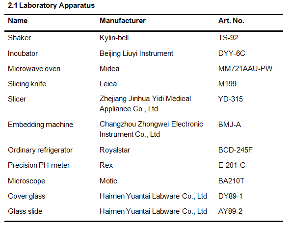 2.1 Laboratory Apparatus
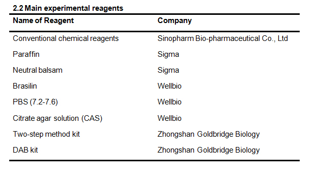 2.2 Main experimental reagents
Kunming (KM) female mice, 8-10 months old, weighing 45-65 g, (animal certificate 43004700021688) and KM female mice, 2-3 months old, weighing 35-40 g, (animal certificate 43004700021689) were used. The mice were provided by the Hunan SLAC Laboratory Animal Co., Ltd, under permit No. SCXK (Hunan) 2011-0003.
Twenty procreated KM female mice (8-10 months old) and five childless KM mice (8 weeks old) were selected. The twenty adult mice were divided into three drug concentration level groups (high, medium, and low) and a control group via a completely randomized block method. Five childless mice were set as the young group. The mice in drug groups were fed Kuntai capsules with a daily dosage of 180 mg/100 g, 90 mg/100 g, and 45 mg/100 g, respectively, once a day, for successive nine days. The mice in control group and the young group were provided with an equal volume of distilled water every day. They were killed an hour after the last administration. They were then laparotomized and their uteri removed. The uteri were saline washed and fixed in a 10 % formaldehyde solution and stored in the refrigerator at 4°C.
3.2 Specimen PreparationFixation: The female mice uteri were fixed in a 10 % formaldehyde solution for 24 hours.
Dehydration: The fixed uteri were washed in tap water and dehydrated using an alcohol gradient method: 30 minutes in 70 % alcohol; 30 minutes in 80 % alcohol; 30 minutes in 90 % alcohol; and, 30 minutes in 100 % alcohol.
Transparency: Post-dehydration, the uteri were placed in a mixture (ratio 1:1) of xylene and 100 % alcohol for 30 minutes; then in xylene-I for 30 minutes; and, finally in xylene-II for 30 minutes until the tissues were transparent.
Paraffining: The transparent uteri tissues were placed in a paraffin cup I for an hour; paraffin cup II for an hour, and paraffin cup III for an hour. The entire process finished in the paraffin melter at a maintained temperature of 57°C.
Embedding: The paraffined uterine tissues were placed in a cassette and marked, and then embedded with the melted paraffin. The paraffin blocks were then decanted after the paraffin solidified.
3.3 Immunohistochemistry1) The slices were baked for 30-60 min at 60°C;
2) Slice dewaxing: The slices were twice placed into xylene for 10 min. They were then, for 5 min each, placed into a 100 % ethanol solution, a 95 % ethanol solution, an 85 % ethanol solution, and a 75 % ethanol solution. Finally, they were immersed in distilled water for 5 min;
3) Heat repair antigen: The slices were immersed into a 0.01M citrate buffer (pH 6.0), and an electric furnace, or microwave oven, heated them to boiling and stopped. The heating was repeated for 6 times after an 8 min. interval. After cooling, they were washed three times in 0.01 M PBS (pH 7.2-7.6) for 3 min;
4) They were then dropped in a 3 % H2O2 for 10 min at room temperature to inactivate endogenous enzymes and then washed three times in PBS for 3 min;
5) Primary antibody incubation: They were dropped in an appropriately-diluted primary antibody (PCNA, LIF), and kept overnight at 4°C. They were then washed three times in PBS for 5 min;
6) Secondary antibody incubation: They were dropped in 50-100 μL anti-rabbit, rabbit IgG antibody - HRP polymer, incubated for 30 min at 37°C and washed three in PBS for 5 min;
7) DAB developing: They were dropped into a prepared DAB developer working solution of 50-100 μL, incubated for 1-5 min at room temperature to control the reaction time in a microscope and washed in distilled water;
8) They were then redyed in hematoxylin for 5-10 min, washed in distilled water, and returned to blue in PBS;
9) They were then dehydrate in alcohol at various levels (60-100 %) for 5 min at each level. Upon removal from the alcohol they were twice, for 10 minutes, placed in a xylene solution. The slices were sealed in neutral balsam, and observed microscopically.
3.4 Data analysisAn ordinary computer was used to sample the images.
IPP (Image-Pro-Plus) software was used to analyze the images.
Notes: The average IOD was the ratio between the cumulative optical density of positive expression site and the area of the sample under the visual field (for cyclogram of 400 times); The general average IOD was a positive analysis for the non-pure nuclear expression indicator; The positive rate was the ratio between the number of positive expression cell nuclei and total number of cell nuclei under the visual field (for cyclogram of 400 times); and The general positive rate is the positive analysis of pure nuclear expression indicator.
3.5 Indicator-related experimental conditions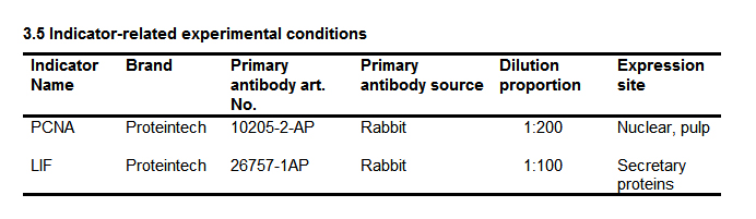 3.5 Indicator-related experimental conditions
A Control
 Fig. 1A Expression of PCNA Proteins by Group: Control
B Low concentration
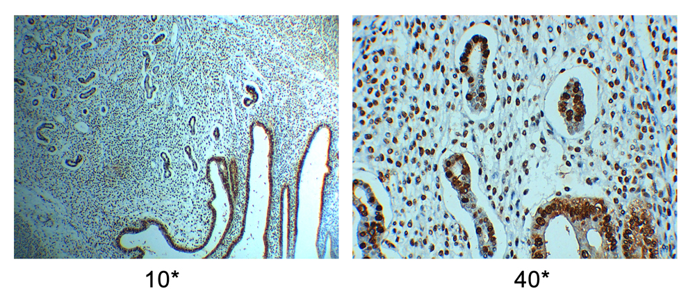 Fig. 1B Expression of PCNA Proteins by Group: Low concentration
C Medium concentration
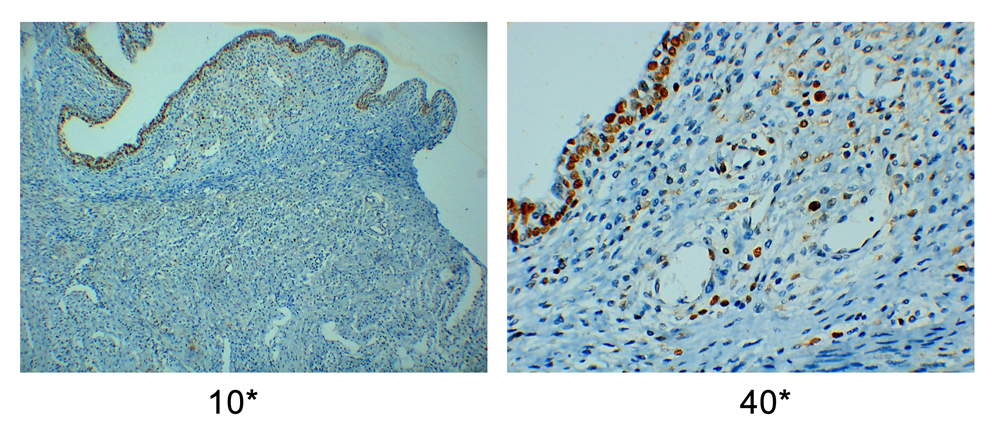 Fig. 1C Expression of PCNA Proteins by Group: Medium concentration
D High concentration
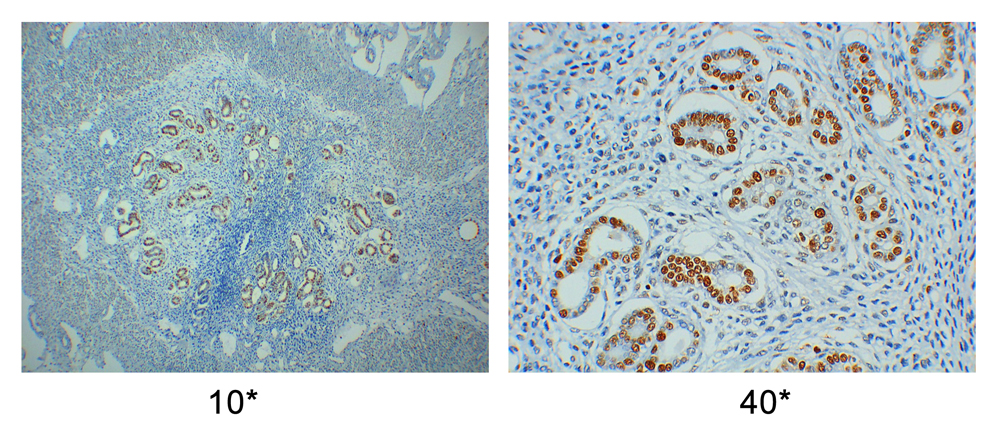 Fig. 1D Expression of PCNA Proteins by Group: High concentration
E Young
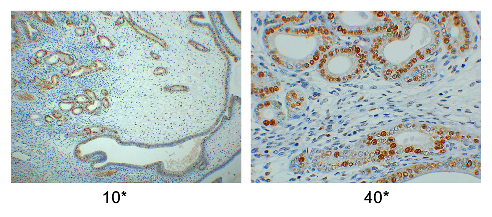 Fig. 1E Expression of PCNA Proteins by Group: Young
A Control
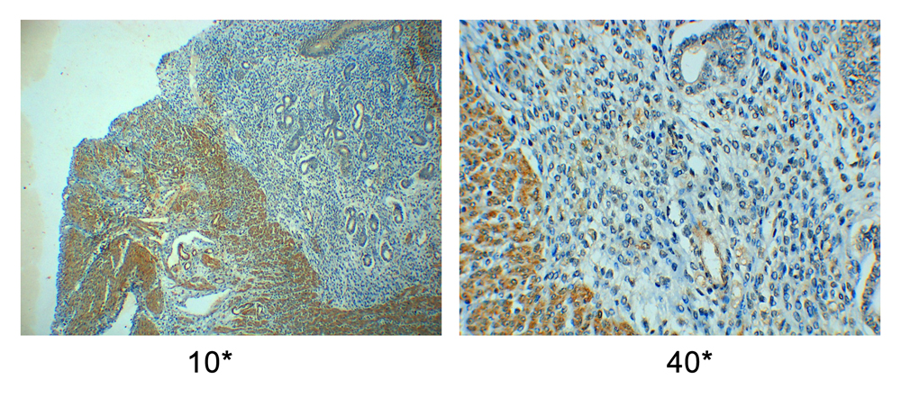 Fig. 2A LIF Protein Expression by group: Control
B Low concentration
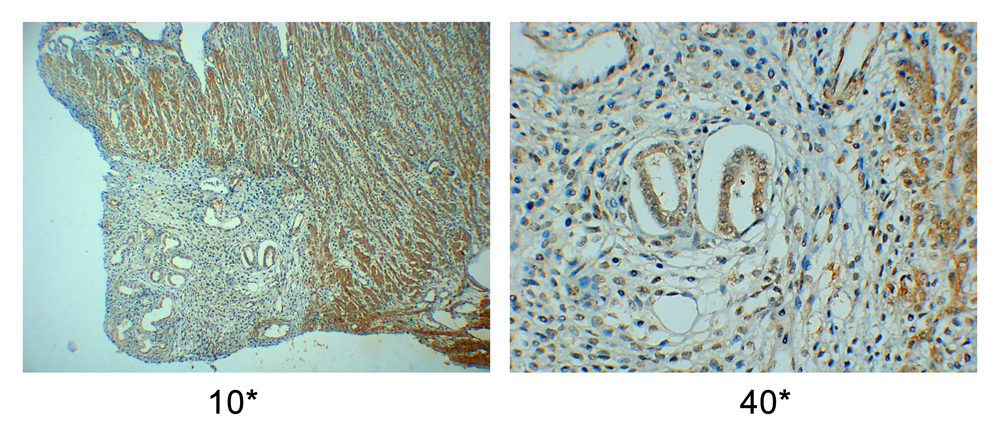 Fig. 2B LIF Protein Expression by group: Low concentration
C Medium concentration
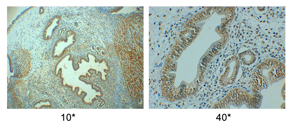 Fig. 2C LIF Protein Expression by group: Medium concentration
D High concentration
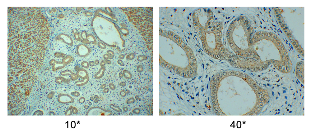 Fig. 2D LIF Protein Expression by group: High concentration
E Young
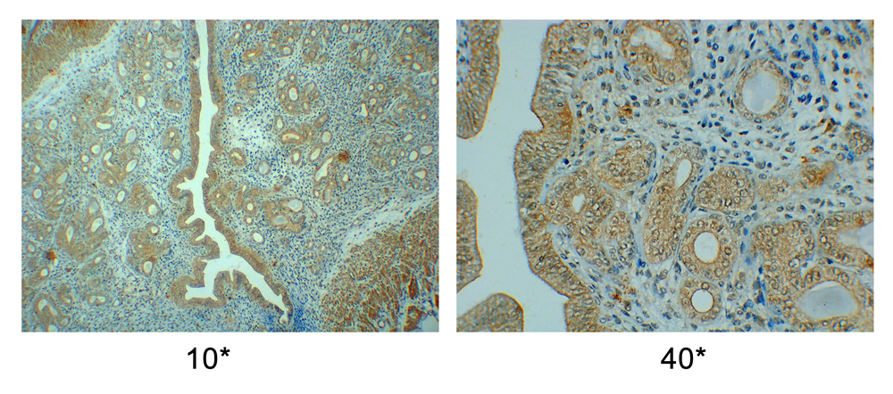 Fig. 2E LIF Protein Expression by group: Young
PCNA and LIF protein expressing in adult female mice decreased compared to young female mice.
Kuntai capsules contain rehmannia glutinosa, radix rehmanniae, rhizoma coptidis, coptis, radix scutellariae, and poria cocos. The contents nourish Yin to lessen fire, regulate Yin and Yang, and sooth nerves and can relieve restlessness. It can tackle symptoms and improve the menopausal syndrome [4]. Some studies [5] show that Kuntai capsules play a two-way regulatory role in hormone level of the menopausal hormonal levels of female mice, and may improve perimenopausal syndrome under conditions of no change of its endogenous hormone level changes.
5.2 Kuntai capsule effects on PCNA protein expression in perimenopausal female miceRecent studies show that PCNA is a marker reflecting cell proliferation activities and plays an important role in DNA replication as an auxiliary protein of DNA polymerase δ. It is an acidic nuclear protein with a molecular weight of 36KD [6]. Jeff R, et al. found that the PCNA gene was subject to up-regulated expression in periimplantation endometriums. It is speculated that PCNA is involved in endometrium proliferation and endometrial decidualization during germ cell adhesion and invasion into the endometrium [7]. This study suggests PCNA protein expression in control, and drug, group endometriums was significantly lower than in young group. This indicates that, during perimenopause, PCNA protein expression in the endometrium is affected. After Kuntai capsule administration, PCNA protein expression in mice endometrium increased significantly suggesting that the contents of Kuntai capsules may significantly alleviate negative effects on the perimenopausal endometrium. This effect also increased with Kuntai capsule dosages.
5.3 Kuntai capsule effects on LIF protein expression in permenopausal female miceLIF is a multi-functional cytokine in the interleukin-6 (IL-6) family with many biological functions and has different biological activities in different tissues [8]. Recently, studies LIF studies have focused on its role in assisting endometrium development which facilitates embryo implantation [9] which improves the infertile patient pregnancy rates. This study suggests that the expression of LIF proteins expression in the control, and drug, group endometriums was significantly lower than that in the young group. This suggests perimenopause may LIF protein expression in the endometrium. After Kuntai capsule administration, LIF protein expression in mice endometrium increased significantly, suggesting that Kuntai capsules may significantly improve the negative effect on perimenopausal endometriums. This improvement also increased with Kuntai capsule dosage increases.
PCNA and LIF protein expression in adult female mice decreased compared to young female mice. The ingredients of Kuntai capsules may significantly improve PCNA and LIF protein expression in female perimenopausal mice. Increased dosages of Kuntain capsules may even more significant improvement.
No conflict of interest and financial disclosure is present. Qiliang Zhou contributed the manuscript, Youru Zhou contributed experimental data and Zhihuo Liu provided language editing services.
This work was supported by a grant from Scientific Research Fund of Hunan Provincial Department of Education (program No.: 15C0158).
1.
2.
3.
4.
5.
6.
7.
8.
9.
Zhou Q, Zhou Y, Liu Z. The Effects of the Ingredients of Kuntai Capsules on LIF and PCNA Protein Expressions in Perimenopausal Mice. Med One. 2017 Apr 25; 2: e170009. https://doi.org/10.20900/mo.20170009

Copyright © 2020 Hapres Co., Ltd. Privacy Policy | Terms and Conditions