Location:Home >> Detail
Med One. 2016; 1(4): 4; https://doi.org/10.20900/mo.20160017

1 Department of Orthopedics, Affiliated Hospital of Hunan Academy of Traditional Chinese Medicine, Changsha, Hunan 410006, P.R. China
2 Department of Anatomy, Changsha Medical University, Changsha, Hunan 410219, P.R. China
3 Department of Clinical Laboratory, Changsha Stomatological Hospital, Changsha, Hunan 410004, P.R. China
First Author: Hui Xu, Associate Chief Physician, Affiliated Hospital of Hunan Academy of Traditional Chinese Medicine.
*Corresponding Author: Jie Luo, Department of Clinical Laboratory, Changsha Stomatological Hospital, Hunan410004, P.R. China.
Objective: To investigate any correlations between TGF-β1-509C/T gene polymorphisms and primary knee osteoarthritis (PKOA) to obtain new new understanding PKOA pathogenesis and new methods for the diagnosis, and treatment of PKOA.
Methods: Eighty-eight PKOA patients and 89 healthy controls were randomly selected from the Department of Orthopedics, the Affiliated Hospital of Hunan Academy of Traditional Chinese Medicine. Polymerase chain reaction- restriction fragment length polymorphism (PCR-RFLP) analysis was performed to test TGF-β1-509 locus genes and genotypes of the subjects. Genotypes and allele frequencies were compared using the chi-square test. Relative risk is represented with odds ratio (OR) and 95 % confidence interval (CI).
Results: The distribution of TGF-β1-509 genotypes for PKOA and healthy control groups complied with Hardy-Weinberg equilibrium law, indicating that a representative population was used. Compared to the healthy control group, the PKOA group had distribution differences for both genotypes and allele frequencies. PKOA group CC genotypic frequency was significantly higher than the healthy control group (44.3 % vs. 13.5 %, χ2 = 20.51, p = 0.0000); allele frequency was significantly higher than the control group (65.9 % vs. 41.6 %, χ2 = 21.08, p = 0.0000). The differences were statistically significant (p < 0.05). No statistically significant differences were found between mild and severe CC genotype frequencies (39.5 % vs. 48.9 %, χ2 = 0.792, p = 0.37), and between C allele frequencies (62.8 % vs. 68.9 %, χ2 = 0.728, p = 0.39) in PKOA group.
Conclusion: TGF-β1-509C/T gene polymorphism associates PKOA in Hunan region patients. allele C carriers in the locus may have higher susceptibiliy to PKOA.
Primary knee osteoarthritis (PKOA) is a common and frequently occurring disease in osteoarthropathic surgeries. Its incidence tends to constantly increase. It has become a chronic disease that seriously harms the health of the middle-aged and elderly [1]. Its etiology, mechanisms, prevention, and treatment has been intensively studied. The pathogenic mechanisms remain unclear and onset is not easily detected [2]. PKOA generation and progression is caused by the synergy of several factors including: local, genetic, environment, and others [3, 4]. This study focuses on correlations between TGF-β1-509C/T polymorphisms and Hunan region PKOA patients to open up a new field for the treatment of PKOA.
Eighty-eight PKOA patients (43 mild, and 45 severe, cases) who had been diagnosed either as outpatients, or, inpatients at the Department of Orthopedics of the Affiliated Hospital of the Hunan Academy of Traditional Chinese Medicine between May 2013 and September 2014 were selected randomly. There was no consanguinity. It included 32 male, and 56 female, patients, aged 47.5 ± 8.3 years old. Inclusion criteria were: 1) Patients with clinical symptoms meeing the Guide of Chinese Orthopaedic Association of Chinese Medical Association on the Diagnosis and Treatment of Osteoarthritis (2007) standards. The Kellgren-Lawrence (K-L) X-ray five level grading criteria for osteoarthritis was employed [5]. Using clinical symptoms in combination with knee x-ray K-L criteria, meeting levels 0, I, or II standards were typed as mild PKOA; levels III, or IV, were typed as severe PKOA; 2) Patients without secondary knee osteoarthritis; and, 3) Patients without hypertension and diabetes and liver and kidney dysfunction. Eighty-nine patients who, afer a physical examination, manifested no PKOA clinical symptoms physical and knee x-ray examination shows no abnormalitites, during same period (May 2013-September 2014) were selected randomly as the healthy control group. This 28 male, and 61 female, patients, aged 48.8 ± 7.5 years old.
2.2 Reagents and InstrumentsThe following reagents and instruments were used in this study: Taq DNA polymerase 5 U/μL (Dongsheng Biotech Co., Ltd, Guangzhou, Guandon, PRC), restriction endonuclease Bsu 500 U/mL (MBI Fermenas ), whole blood genomic extraction kit, DNAgel extraction kit (Guangzhou Dongsheng Biotech Co., Ltd), PCR instrument (ABI corporation ), electrophoresis (Beijing Liuyi Biotechnology Co., Ltd), and integrated gel imaging analysis system (Beijing Sage Creation Science Co., Ltd).
2.3 Gene Polymorphisms TestingFasting, peripheral venous blood samples of 2 ml were taken from each subject in the early morning. The samples were treated with an anticoagulant EDTA-Na2 and stored at 4°С. Genomic DNA was extracted within a week. If genomic DNA was not extracted within a week, the samples were stored at -20°С. Genomic DNA extraction from the peripheral bleed was performed in accordance manufacturer’s instructions. Gene sequences were found in the NCBI database (rs1800469). The PCR primer was designed using software Primer 5.0 (sequence of forward primer: 5'-GCTACGGCGTGGAGTGCTGA-3'; sequence of reverse primer: 5'-AGAGGACCAGGCGGAGAAGG-3') [6].
Primer sequences were synthesized by the BGI Gene Research Center. Amplified fragment length was 506 bp. PCR reaction conditions were pre-degenerated at 94°С for 5 min, followed by 35 cycles at 94°С, 30 sec., 56°С 30 sec., and 72°С 30 sec., and finally, 72°С 5 min and stored at 4°С. Amplified products were digested via endonuclease Bsu36I (Eco81L). The digested products were 10g/L agarose gel electrophoresis.
2.4 Statistical AnalysisThe statistical analysis was performed by statistical software SPSS13.0. The Hardy-Weinberg equilibrium, genotype, and allele, frequencies were compared using the chi -square test and relative risk was represented with the odds ratio (OR) and 95 % confidence interval (CI). p < 0.05 indicates a statistically significant difference.
If -509 locus (-CTTC-) existed in the amplified fragments, the interchange between thymine (T) and cytosine (C) might appear at the -509 locus (-CTTC-), a Bsu36l endonuclease locus would be generated, and produce two fragments: 329 bp, and 177 bp after Bsu36l endonuclease digestion. TT geneotype had only one fragment (506 bp), CC genotype had two fragments (329 bp and 177 bp), and CT genotype had 3 fragments (506 bp, 329 bp and 177 bp) (Fig. 1).
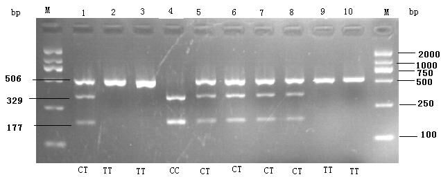 Fig. 1 RFLP Electrophoresis
Three genotypes were selected randomly and sequenced. The sequenced results were completely consistent with the electrophoresis results (Fig. 2).
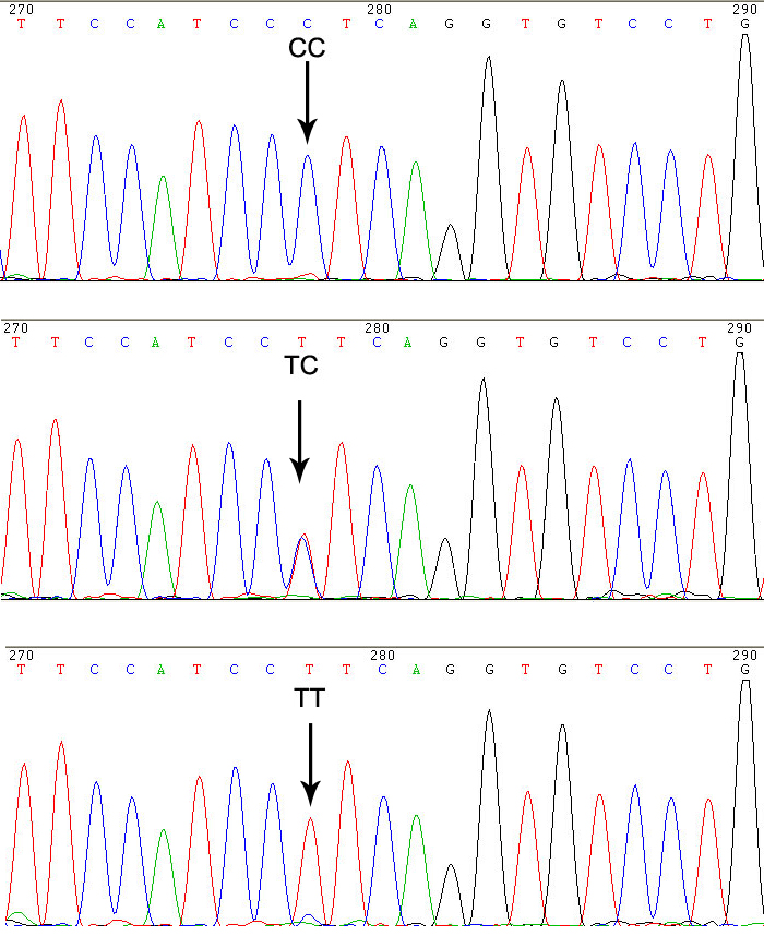 Fig. 2 TGF-β1-509 Polymorphic Locus Sequencing
Testing showed that the distribution of TGF-β1-509 genotypes in both the PKOA, and the control, groups complied with the Hardy-Weinberg equilibrium law (χ2 = 0.135, p = 0.934; χ2 = 2.190, p = 0.335, p > 0.05), indicating that a representative population was used. The PKOA group had a distribution difference between genotype and allele frequency compared to the control group.
PKOA group CC genotypic frequency was significantly higher than the control group (44.3 % vs. 13.5 %, χ2 = 20.51, p = 0.0000). Allele frequency was significantly higher than that of control group (65.9 % vs. 41.6 %, χ2 = 21.08, p = 0.0000) (p < 0.05). The relative risk analysis of allele frequencies showed that C allele carriers risk of PKOA was 2.72 times that of T allele (OR = 2.72, 95 %, CI = 2.57 -2.88, p = 0.0000) (Table 1).
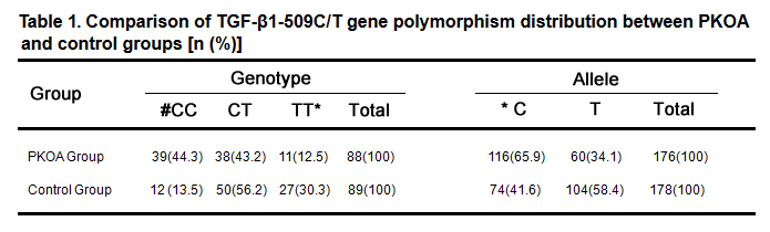 Table 1. Comparison of TGF-β1-509C/T gene polymorphism distribution between PKOA and control groups [n (%)]
Notes: # χ2 = 20.51, p = 0.0000; *χ2 = 21.08, p = 0.0000, OR = 2.72, 95 %, CI = 2.57-2.88, p = 0.0000
No statistical differences were found between mild and severe CC genotype frequencies (39.5 % vs. 48.9 %, χ2 = 0.792, p = 0.37) in PKOA group and between C allele frequencies (62.8 % vs. 68.9 %, χ2 = 0.728, p = 0.39), the differences were not statistically significant (Table 2).
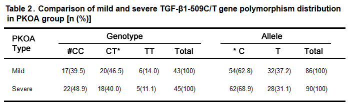 Table 2. Comparison of mild and severe TGF-β1-509C/T gene polymorphism distribution in PKOA group [n (%)]
Notes: #χ2 = 0.792, p = 0.37; *χ2 = 0.728, p = 0.39
OA incidence is a complicated, pathological process. Current PKOA research focuses on cartilage and synovium. Cytokines affecting cartilage, and synovial, cells include growth factors, chemokines, interleukins, and tumor necrosis factors. TGF-β plays an important role in OA osteophytes and synovial hyperplasia. TGF-β1 is in the TGF-β superfamily and plays an important role in cartilage growth and reconstruction. It is an important PKOA-associated gene. Studies have shown an association between the single nucleotide polymorphism (SNP) locus of the TGF-β1 gene and OA. But research results are not uniform. Youcheng Wang [7] found that the TGF-β1 C1348-T locus polymorphism significantly associated with PKOA. A common variation in the population of China occurs at -509C/T, and correlation studies of the -509C/T locus and PKOA have not been reported outside China.
A correlation analysis was performed in this study. The subjects were two populations selected from a PKOA group and a control group, with the same, or very similar, genetic makeup. The purpose was to compare allele frequency differences in certain candidate gene polymorphisms between the two populations. Correlation analyses should show that the allele frequency distribution in the PKOA group will be significantly higher than that in the control group, if certain alleles, or its adjacent locus, are associated with disease susceptibility. PKOA group allele frequency should be significantly lower than that the control group if it relates to disease resistance and protection. Experimental results showed genotypic frequency, and allele distribution, differences between the PKOA group and healthy controls. CC genotypic frequency in the PKOA group was higher than that in the control group (44.3 % vs. 13.5 %). The C allele frequency was higher than in the control group (65.9 % vs. 41.6 %), with the difference being statistically significant (p < 0.05). Logistical regression analysis results showed that, compared to the allele T-carrying population, the allele C-carrying population risk for PKOA is 2.72 times greater (OR = 2.72, 95 %, CI = 2.57 - 2.88). This suggests that the -509 allele C in the TGF- β1 promoter region may be a PKOA involved gene and that allele T may be a protective gene. This may relate to: why the change of allele C towards allele T increased TGF- β1 translation, and expression, levels; inhibited immune cell activation; and, increased osteoarthritis resistance [8]; or why greater TGF-β1 expression mediated cartilage synthesis; inhibited collagen, and proteoglycans, decomposition; protected the cartilage matrix from hydrolyzation, and protease, damage and achieved cartilage damage reversal [9, 10].
Jing Guo, et al. [11] found that cartilage, and synovial, cell apoptosis in the OA group negatively correlated with TGF-β1 expression, high TGF-β1 high expression in the OA group may negatively regulate cartilage, and synovial, cell apoptosis generation, and delay osteoarthritis onset and progression. Ming Chen, et al.. [12] found that knee osteoarthritis (KOA) patient synovial fluid TGF-β1 levels were significantly negatively correlated with KOA stages. KOA patient synovial fluid TGF-β1 levels at the middle and advanced stages was significantly lower than that in the control group (p < 0.05). Compared to the healthy control group, early-stage KOA patient synovial fluid TGF-β1 levels werenot statistically significant (p < 0.05). TGF-β1 might protect, and repair, osteoarthritis and it was a measure for disease severity. This study divided PKOA was into mild and severe. The experiments concluded that the differences were not statistically significant comparing genotype and allele frequency of the two groups. The -509C/T gene polymorphism was not associated with PKOA severity.
The differences in the findings may relate to the sample quantity and size, different regions, ethic groups, and studied-population genetic heterogeneity PKOA is a complicated pathological process involving multi-gene inheritance. Genetic heterogeneity, environmental factors, synergy between genetic factors, and other complicated factors might affect PKOA and warrant further study.
This work was supported by grants from the Science & Technology Program of the Department of Science and Technology of Hunan Province (program number: 2013FJ3152) and the program of Hunan Academy of Traditional Chinese Medicine (program number: 201224).
The authors declare no conflict of interests.
1.
2.
3.
4.
5.
6.
7.
8.
9.
10.
11.
12.
Xu H, Zhou Q, Luo J. Correlation Between TGF-β1-509C/T Gene Polymorphisms and Primary Knee Osteoarthritis-A Clinical Study. Med One. 2016; 1(4): 4; https://doi.org/10.20900/mo.20160017

Copyright © 2020 Hapres Co., Ltd. Privacy Policy | Terms and Conditions