Location: Home >> Detail
Regen Med Front. 2020;2(1):e200002. https://doi.org/10.20900/rmf20200002

Orthopaedic Research Center, C. Wayne McIlwraith Translational Medicine Institute, Department of Clinical Sciences, Colorado State University, Fort Collins, CO 80523, USA; Tel.: +1-970-297-5071
Culture-expansion is a common step in the use of autologous chondrocytes for cartilage tissue engineering. Chondrocytes dedifferentiate in monolayer expansion culture, and conditions that are sufficient to induce redifferentiation change as a function of cumulative growth. The objective of this review was to characterize the relationship between expansion and redifferentiation, from which requirements to induce redifferentiation were identified for selected approaches to cartilage tissue engineering. While chondrocytes dedifferentiate rapidly in expansion culture, transferring the cells to a three-dimensional scaffold is sufficient to induce redifferentiation for up to ~6 population doublings (PDs). Redifferentiation is possible beyond 6 PDs, although exposure to chondrogenic cytokines is needed. These data indicate that dedifferentiation with expansion for treating focal defects (~6 PDs) can be reversed with transfer to a scaffold, while joint resurfacing (~10 PDs) is anticipated to require exposure to chondrogenic cytokines. During expansion, growth factor supplementation can accelerate proliferation and improve redifferentiation. However, redifferentiation may require chondrogenic cytokines, and should be considered for approaches that involve expansion beyond the limit for spontaneous redifferentiation.
Culture-expansion is often necessary for tissue engineering therapies as the number of cells needed to fill areas of damaged or diseased tissues typically exceeds the availability of primary cells. Cartilage tissue engineering has been enabled by the ability of articular chondrocytes to readily proliferate in monolayer culture using standard cell culture medium. However, chondrocyte expansion is associated with loss of the native phenotype, which must be restored after expansion as implanted chondrocytes are expected to secrete and assemble neo-cartilage. Chondrocyte dedifferentiation with expansion, and redifferentiation after, has been the focus of many studies over approximately four decades. These data have revealed that chondrocyte dedifferentiation progresses with time in expansion culture. Likewise, the conditions that are needed to induce redifferentiation change as a function of culture expansion. Therefore, for tissue engineering strategies it is important to consider how much chondrocytes must be expanded in order to ensure that an effective redifferentiation strategy is in place.
For studies evaluating chondrocyte expansion and redifferentiation, the extent to which chondrocytes have been expanded appears to be largely arbitrary, with few studies targeting expansion needs for specific tissue engineering therapies. Therefore, the primary objective of this review is to characterize chondrocyte expansion for selected approaches to cartilage tissue engineering so that the steps needed to induce redifferentiation can be identified. Expansion for the use of autologous chondrocytes to treat focal cartilage defects or joint resurfacing was calculated in terms of cumulative population doublings (PDs), which is the number of times a starting population of cells must double to reach a target cell number. Across the studies referenced for this review there is a moderate amount of variability in the methods used to promote expansion and redifferentiation. Therefore, we identified techniques that are particular effective in promoting expansion and/or redifferentiation.
The process of expanded cells in monolayer culture involves seeding at a subconfluent density and then detaching the cells from the monolayer surface when the proliferating cells approach confluence. Detaching the cells and reseeding monolayer surfaces at a subconfluent density is known as passaging, and the extent to which a batch of cells has been expanded is often described by the number of passages. However, the amount of cell growth per passage is dependent on the initial seeding density, which for the studies referenced here varied by up to 100-fold. To account for difference in seeding density our analysis was based on cumulative PDs, which were provided or estimated when sufficient information was provided.
Currently, induction of endogenous repair using the microfracture procedure is used to treat small defects up to approximately 3 cm2 in area [1], while larger defects are treated with a form of chondrocyte implantation. For tissue engineering approaches using autologous chondrocytes, documentation of the Autologous Chondrocyte Implantation (ACI) technique provides quantitative values on defect size, autologous tissue harvest and expected chondrocyte yield, and cell seeding density in the defect from which the number of required PDs can be calculated. For defect size, calculations were based on an area of 6.5 cm2, which is the average size reported across more than 100 patients that were treated with ACI [2]. In the same study, chondrocytes were implanted at 1–2 × 10⁶ per cm2 of surface area, which is consistent with other techniques for filling cartilage defects with cells [3]. Given that adult human knee cartilage is approximately 2 mm thick [4], the density of the implanted cells is approximately 7.5 × 106 cm−3, which is comparable to chondrocytes in human cartilage [5]. Assuming cell seeding of 1.5 × 106 cm−2, the total number of cells for a 6.5 cm2 defect is 9 × 10⁶. For ACI approximately 200–300 mg of cartilage is harvested [6], from which approximately 0.25 × 10⁶ cells are obtained [6,7]. Therefore, it is assumed that autologous chondrocytes must be expanded by 40-fold, or approximately 5.3 PDs. It should be noted that deviation from the mean parameters is likely to change the number of PDs by no more than 2. For example, from Neimeyer et al., the standard deviation of the defect size ranged between 2.5 and 10.5 cm2 [2]. For this range of defect sizes the number of PDs needed from a standard cartilage biopsy is 3.9 and 6.5, respectively. Similarly, variation in the number of cells obtained among patients is encompassed by approximately a single PD. For example, in Brittberg et al., the yield of primary chondrocytes ranged between 0.18 and 0.45 × 10⁶ [8], which in terms of chondrocyte growth is a difference of 1.3 PDs. Taken together, it is assumed that use of autologous chondrocytes to treat focal cartilage defects requires 4–7 PDs.
Osteoarthritis is associated with progressive loss of cartilage, and patients that have experienced widespread cartilage erosion would benefit from approaches to repair large areas or whole surfaces of joints [9,10]. In young adults, the mean knee surface area was reported to be approximately 100 cm2 [11], which is approximately 15-fold larger than the 6.5 cm2 focal defect used to calculate model expansion of autologous chondrocytes above. Assuming a cell yield of 0.25 × 10⁶, and a seeding density of 1.5 × 106 cm−2, autologous chondrocytes would require approximately 10 PDs for resurfacing large areas of cartilage.
Chondrocyte Dedifferentiation in Expansion CultureCartilage differs from other connective tissues in that the collagen content is almost entirely type II. Therefore, researchers have considered loss of type II, and increases in type I collagen expression as indicators of dedifferentiation (Table 1). In this section, the cited studies did not report the number of PDs associated with changes in collagen expression, and in all but one an estimate could not be performed due to lack of data. While chondrocyte growth rates can vary greatly among studies it is likely that proliferation in these studies was on the order of 2–3 PDs per week. In 1976, Mayne et al. considered collagen synthesis by chick embryo chondrocytes in expansion culture [12]. Serial passaging of chick embryo chondrocytes selected via colony-formation resulted in a transition in collagen synthesis. While four passages were needed before collagen synthesis resembled that of control fibroblast cultures, the largest decline in type II collagen synthesis occurred between the first and second passage. Similar trends reported for rabbit articular chondrocytes [13]. These data indicated that chondrocyte dedifferentiation is progressive with expansion, but is initiated rapidly. This conclusion is supported by subsequent studies evaluating type I and type II collagen gene expression. For adult human chondrocytes, the ratio of type II to type I gene expression decreased by more than 10-fold over the first two weeks in expansion culture, with more graduate decreases over two subsequent weeks [14]. Expansion of chondrocytes from adult human cartilage biopsied from diseased joints led to an approximate 400-fold decrease in the ratio of type II/type I gene expression over three weeks [7]. In Barlic et al., collagen gene expression of adult human chondrocytes was evaluated over the first 10 days of expansion culture [15]. Type II collagen gene expression decreased ~5-fold, while type I gene expression increased ~300-fold. In Darling et al., young adult goat chondrocytes were evaluate for collagen gene expression after 4 passages [16]. The cells were subculture at a ratio of approximately 1:3 which translates to approximately 1.6 PDs per passage. Compared to primary cells, type II collagen gene expression decreased approximately 3-fold at the first passage, and nearly 10-fold after 4 passages (approximately 6 cumulative PDs). Type I collagen gene expression increased approximately 20-fold across 4 passages. In Jakob et al., expansion of human chondrocytes through 5–7 PDs decrease the ratio of type II/type I collagen gene expression by more than 300-fold [17]. These data indicate that dedifferentiation is well-established for the tissue engineering needs as defined above.
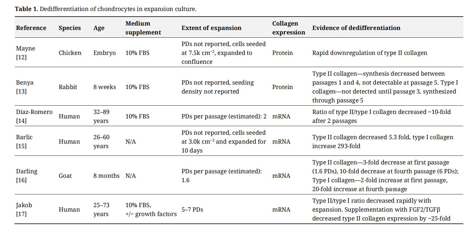 Table 1. Dedifferentiation of chondrocytes in expansion culture.
In 1982, Benya and Shaffer investigated the potential to induce redifferentiation of expanded lapine chondrocytes by transferring the cells from adherent monolayer to suspension culture, which was achieved by encapsulating the cells in agarose hydrogel [18]. Chondrocytes were subcultured at a ratio of 1:3, therefore it is assumed that at each passage the cells proliferated through 1.6 PDs. Chondrocytes were transferred to agarose at the fourth passage (6.4 PDs), a point at which dedifferentiation was indicated by type I collagen synthesis, and suppressed proteoglycan and collagen synthesis in monolayer. After suspension in agarose, rapid redifferentiation was indicated by re-expression of type II collagen by day 2, and increasing proteoglycan and collagen synthesis that reached levels comparable to primary cultures by day 10. Given that the same culture medium was used for expansion and redifferentiation, the authors concluded that three-dimensional culture was sufficient to induce redifferentiation. These data indicate that despite robust dedifferentiation in monolayer, three-dimensional culture was sufficient to restore the native phenotype. Subsequently, redifferentiation of expanded chondrocytes in vitro was demonstrated in various scaffolds such as alginate hydrogel [19] or synthetic polymer [20]. Therefore, from a tissue engineering perspective expanded chondrocytes can be expected to spontaneously redifferentiate in that the fundamental step of seeding cells in a scaffold can reverse dedifferentiation.
Limits in the Ability of Expanded Chondrocytes to RedifferentiateIn Benya and Shaffer, expansion between passage 4 (6.4 PDs) and 6 (9.6 PDs) resulted in a trend of decreasing collagen synthesis in agarose culture, which the authors raised as a potential indicator of diminishing redifferentiation with extensive expansion [18]. Subsequent in vivo studies confirmed that the ability of chondrocytes to redifferentiate strictly with transfer to a three-dimensional environment is lost as a function of growth. In Passaretti et al., redifferentiation of porcine chondrocytes as a function of passage number (estimated to be 4–5 PDs per passage) was tested using a subcutaneous mouse model [21]. As determined by histology, cartilage was produced by unexpanded chondrocytes as well as chondrocytes expanded to passage 1, although chondrocytes that were expanded beyond passage 1 did not synthesize cartilage. In terms of PDs, chondrocytes that were expanded through ~4.5 PDs were capable of spontaneous redifferentiation, while chondrocytes expanded through 9 and higher PDs were not. Similar results were obtained for human chondrocytes that were evaluated for redifferentiation after subcutaneous implantation as cartilage formation was observed for expansion through 2 PDs but not 6 PDs [22]. In Dell’Accio et al., redifferentiation of adult human chondrocytes was evaluated following intramuscular injection in mice [23]. Unexpanded chondrocytes formed cartilage in vivo, while chondrocytes that were expanded through 4–6 PDs did not. Taken together, these data indicate that chondrocytes lose the ability to redifferentiate after approximately 5–6 PDs.
Induction of Redifferentiation Using Chondrogenic Growth FactorsIn the laboratory studies cited above, redifferentiation of expanded chondrocytes was evaluated in culture medium that was the same as or closely resembled medium use for expansion. The use of similar media was intended to rigorously test the ability of chondrocytes to redifferentiate based on transfer from monolayer to three-dimensional culture. However, subsequent studies reported that growth factors from the transforming growth factor beta (TGFβ) superfamily added during three-dimensional culture strongly promote redifferentiation. In many cases, redifferentiation was evaluated in a serum-free formulation containing TGFβ that induces robust chondrogenesis of MSCs [24], which will be referred to as chondrogenic medium. Scaffold-free pellets of expanded adult human chondrocytes that were salvaged from cell preparations for ACI and cultured in chondrogenic medium showed higher levels of redifferentiation compared to cultures maintained in medium supplemented with 10% serum [25]. Similar results were reported for adult human chondrocytes expanded through ~4 PDs [26]. Importantly, numerous studies have demonstrated redifferentiation of extensively-expanded chondrocytes with growth factor supplementation. For example, Yaeger et al. evaluated adult human chondrocyte redifferentiation after expansion through 12 PDs [27]. Expanded chondrocytes that were cultured with TGFβ1 re-expressed type II collagen, whereas samples cultured in the absence of TGFβ1 did not. In Bianchi et al., chondrocytes from human osteoarthritic cartilage that were expanded through approximately 9 PDs accumulated cartilage-like matrix in chondrogenic medium, while control samples cultured in chondrogenic medium in which TGFβ3 was withheld accumulated only a small amount of cartilage matrix [28]. These data are consistent with the concept that spontaneous redifferentiation is lost after approximately 6 PDs. In Ono et al., adult human chondrocytes were evaluated for redifferentiation in culture medium containing TGFβ3 and bone morphogenetic protein 2 as a function of passage number (~2.3 PDs/passage) [29]. Robust redifferentiation was observed up to passage 4 (~9 PDs), with comparable gene expression and matrix accumulation among the 4 passages. Additional studies have demonstrated redifferentiation in chondrogenic medium for adult human chondrocytes expanded through 8–9 PDs [30,31]. For non-human chondrocytes, similar patterns of redifferentiation with chondrogenic medium have been reported for bovine [32], canine [33], and lapine [34] chondrocytes. These data demonstrate redifferentiation of chondrocytes expanded beyond 6 PDs is possible with exposure to chondrogenic growth factors (Table 2).
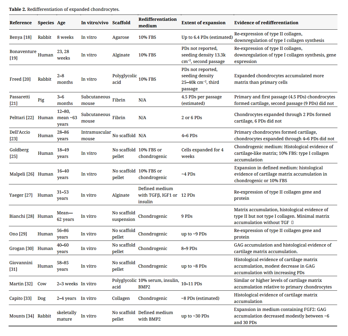 Table 2. Redifferentiation of expanded chondrocytes.
Tissue engineering strategies can be designed around ex vivo manipulation beyond expansion to generate matrix-rich cell-seeded grafts for implantation. However, it is much more common to obtain cells, combine with a scaffold, and then implant with the expectation that the body provides supportive cues for tissue regeneration as is performed for ACI. While chondrocyte dedifferentiation occurs almost immediately after seeding into expansion culture, transferring to a carrier scaffold can induce redifferentiation for up to approximately 6 PDs. Therefore, isolating chondrocytes from ~300 mg of autologous cartilage for focal defects is within, but likely approaching the limit for spontaneous redifferentiation. Expansion of autologous chondrocytes for larger areas of cartilage is expected to exceed the limit for spontaneous redifferentiation. Instead, providing chondrogenic cues appears necessary to induce redifferentiation of extensively-expanded chondrocytes. In this manner extensively-expanded chondrocytes resemble progenitor cells that do not undergo spontaneous chondrogenesis in vitro [24], and have been paired with strategies to induce differentiation before implantation [35,36]. To date, in experimental animal models the effectiveness of ex vivo, growth factor-induced redifferentiation on cartilage matrix accumulation in vivo has been mixed. Prior to subcutaneous implantation, adult human chondrocytes expanded through 8–10 PDs that were cultured in chondrogenic medium acquired significantly more cartilage matrix compared to cells cultured in expansion medium [37]. In experimental focal cartilage defects, modest to no benefit was reported for pre-implantation chondrogenic culture relative to acellular controls in a goat model [38], and chondrocytes cultured in the absence of TGFβ in a rabbit model [39]. More favorable results were reported for expanded adult canine chondrocytes as cell-seeded grafts that were cultured in chondrogenic medium for approximately 4 weeks remained cartilage-like 12 weeks after implantation [40].
Culture Conditions that Are Favorable for Chondrocyte Expansion and RedifferentiationLaboratory studies have studied the biology of chondrocytes in a wide range of culture conditions. In this section we consider factors that appear to be particularly supportive of chondrocyte expansion and redifferentiation.
Expansion culture medium—basal medium
From the studies cited in this review, basal media used for culture expansion was Dulbecco’s modified Eagle’s medium (DMEM), Ham’s F12 (F12), or a mixture of DMEM and F12. In most cases it was not stated whether the glucose concentration in DMEM was high (4.5 g/L) or low (1 g/L). Among the studies it was not clear that the choice of basal medium influenced the rate of proliferation (most commonly a population doubling every 2–3 days) or subsequent redifferentiation. However, from Jiang et al., low glucose medium may be favorable for maximizing the growth rate of chondrocytes [41].
Growth factor supplementation
In the cited studies the most common medium supplement was fetal bovine serum alone. However, additional supplementation with growth factor can significantly enhance the rate of proliferation. Fibroblast growth factor 2 (FGF2) has been identified as a particular effective mitogen. The stimulatory effect of FGF on rabbit auricular chondrocyte proliferation was reported by Gospodarowicz and Mescher in 1977 [42]. For adult rabbit articular chondrocytes FGF supported rapid and nearly linear growth as a function of time in culture up to 20 PDs, while proliferation in control medium without FGF plateaued at fewer than 10 PDs [34]. Similarly FGF2 reduced the population doubling time by approximately 50% for sheep and human auricular chondrocytes [43], and modestly for immature bovine chondrocytes [44]. Importantly, expansion with FGF2 can improve the propensity for chondrocyte redifferentiation. Adult human chondrocytes expanded through 8–9 PDs resulted in greater histologic evidence of redifferentiation in chondrogenic medium when expanded in FGF2 [45]. Immature bovine chondrocytes expanded through ~11 PDs in FGF2 redifferentiated in expansion medium to a similar extent as primary cells, while redifferentiation was much less robust for chondrocytes expanded without FGF2 [44]. For expanded adult rabbit chondrocytes in chondrogenic medium, matrix accumulation for cells expanded with FGF2 was higher than cells cultured in the absence of FGF2 for the same number of PDs [34]. FGF2 has been used in combination with TGFβ [17], TGFβ and platelet-derived growth factor [33,46], and TGFβ and epidermal growth factor in serum-free medium [26,31]. In each case, medium containing growth factors supported a higher growth rate and/or increased propensity for redifferentiation in chondrogenic medium compared to chondrocytes expanded in medium supplemented with serum only. It is important to note that rapid growth and dedifferentiation may compromise the ability of chondrocytes to spontaneously redifferentiate. For example, in Jakob et al., increases in the rate of proliferation with exposure to FGF2 and TGFβ coincided with a severe suppression of type II collagen expression in expansion culture, and subsequently poor redifferentiation in the absence of chondrogenic factors [17].
Effect of insulin on redifferentiation
Insulin is a component of a commonly-used medium supplement (insulin-transferrin-selenium (ITS)) that was developed to support cells in low serum or serum-free medium. ITS is often used in medium to redifferentiate chondrocytes with TGFβ, but is typically not controlled for as a chondrogenic factor. However, several studies have indicated an important role of insulin in chondrocyte redifferentiation. For example, in Yaeger et al., redifferentiation of human chondrocytes expanded through ~12 PDs was evaluated as a function of insulin and TGFβ exposure [27]. Type II collagen gene expression was induced by medium containing insulin and TGFβ. Conversely, gene expression of type II collagen was absent for cultures maintained in medium supplemented with TGFβ alone. In Ahmed et al., redifferentiation of immature bovine chondrocytes expanded through 7–8 PDs was evaluated as a function of medium components of a serum-free formulation that did not contain TGFβ. Insulin was found to be a critical factor for generating histological evidence of redifferentiation [47]. Similar results have been reported for bone marrow MSCs as withholding insulin or ITS from chondrogenic medium greatly suppressed chondrogenesis [48,49]. As demonstrated for immature bovine chondrocytes, cartilage matrix synthesis by primary chondrocytes can be stimulated by insulin [50]. These data suggest that insulin exposure should be considered an important aspect of chondrocyte redifferentiation for extensively-expanded cells, and possibly as a supporting factor for maximizing matrix production.
Effect of dedifferentiation on cartilage matrix synthesis by redifferentiated chondrocytes
Culture expansion can potentiate cartilage matrix synthesis when redifferentiation is induced using growth factors. For example, expanded chondrocyte have been shown to secrete more cartilage matrix than primary cells in expansion [20] or chondrogenic [28,40,51] medium. Two studies indicated that improved cartilage matrix synthesis with expansion is associated with enhanced dedifferentiation. In Jakob et al., expanding chondrocytes in the presence of FGF2 and TGFβ resulted in a rapid and large (~25-fold) decrease in type II collagen gene expression relative to control cultures expanded without growth factors [17]. Compared to controls, chondrocytes expanded through 5–7 PDs with FGF2/TGFβ expressed higher levels of type II collagen in chondrogenic medium. Similar results were reported by Barbero et al. as decreased type II collagen gene expression with 5–8 PDs was associated with greater cartilage matrix accumulation and type II collagen gene expression in chondrogenic redifferentiation culture [46]. Therefore, enhancing dedifferentiation during expansion should be considered for tissue engineering strategies that include exposure to chondrogenic cues after expansion.
In addition to downregulation of type II collagen expression, a strong propensity for redifferentiation may be indicated by properties associated with progenitor cells. Expanded chondrocytes have been described as progenitor cells based on an ability to undergo multilineage differentiation [41,46,52–54] and expression of cluster of differentiation (CD) antigens associated with human MSCs [14,41,52,53,55]. From these studies, expanded chondrocytes demonstrated a strong propensity for cartilage regeneration as indicated by equivalent or superior matrix accumulation in chondrogenic medium relative to bone marrow MSCs [41,53,55]. While certain progenitor-like CD markers are expressed soon after primary chondrocytes are seeded into monolayer [53,56], a subset that are expressed over time in a manner consistent with loss of type II expression [41,56] may be particularly robust indicators of progression of dedifferentiation.
Effect of Donor Age on Chondrocyte RedifferentiationData for this review were compiled from studies in which donor ages ranged from juvenile to elderly. Given that cartilage tissue engineering is predominantly used for young to middle-aged adults [2], it is important to consider whether young or old chondrocytes reflect this target population. The possibility that aging may negatively affect chondrocyte redifferentiation is indicated by primary chondrocytes, which have shown a moderate to severe decline in matrix synthesis with aging [57–59]. However, it is not clear that a similar trend occurs for expanded chondrocytes. When comparing juvenile and adult chondrocytes, redifferentiation following limited expansion has been reported to be comparable for culture in medium supplemented with 10% FBS or chondrogenic medium [60–62]. In one study, matrix synthesis by unexpanded chondrocytes was much higher for juvenile cells [62], which further indicates that the behavior of primary chondrocytes is not necessarily representative of expanded and redifferentiated cells. Conversely, with extensive expansion juvenile chondrocytes retained a greater capacity for redifferentiation compared to adult cells [61,62]. Additional studies are needed to better define expansion limits within which redifferentiation of juvenile chondrocytes is consistent with adult cells. For the adult population, Barbero et al. evaluated redifferentiation in chondrogenic medium as a function of decade of life [63]. Chondrocytes expanded in medium supplemented with 10% FBS through ~4.5 PDs did not show a statistically significant effect of aging, with only modest differences in mean proteoglycan accumulation from the 20s to 70 and older. For serum-free medium supplemented with growth factors (~8 PDs), mean proteoglycan accumulation decreased by more than 50% between the youngest and oldest age group, although differences among ages were not statistically significant due to high variability among donors. Taken together these data suggest that studies that are based strictly on elderly donors may report redifferentiation that is moderately lower than would be expected for young to middle-aged adult chondrocytes.
Chondrocyte dedifferentiation is an unavoidable consequence of culture-expansion. Dedifferentiation can be reversed even with extensive expansion, although the amount of expansion dictates whether chondrocytes can spontaneously redifferentiate in three-dimensional culture or if additional chondrogenic factors are needed. Autologous chondrocytes for focal cartilage defects can be assumed to redifferentiate with implantation, while resurfacing of large areas of cartilage with autologous cells chondrocytes are expected to require additional chondrogenic cues. For these conclusions there are several limitations to consider. Differences in the behaviors of chondrocytes among species should be considered as demonstrated in Tseng et al. where sheep chondrocytes were not necessarily consistent with human [43]. The effect of extensive expansion should be evaluated in greater detail as in vitro aging may significantly diminish the ability of redifferentiated chondrocytes to secrete neo-cartilage [34]. The studies cited in this review used a wide range of subconfluent seeding densities for expansion cultures, which may impact dedifferentiation as relatively high density slows dedifferentiation, and low density promotes dedifferentiation [41,64]. Trends in chondrocyte expansion and redifferentiation identified in the laboratory may not strictly translate to cartilage regeneration in vivo as indicated by long-term follow up on ACI procedures, which has shown that repair tissue often contains a mix of hyaline and fibrocartilage [65]. For ACI, implanted chondrocytes have been shown to persist over a period of weeks in animal studies, with defects filled with both implanted and endogenous cells [66,67]. Therefore, interactions between chondrocytes and endogenous cells may influence whether repair hyaline or fibrocartilage tissue. While differences in the choice of materials and techniques are likely to persist among laboratories, designing studies around anticipated expansion needs for tissue engineering therapies can help address variability in the propensity of chondrocytes to redifferentiate with expansion. Further, reporting expansion in terms of PDs is recommended to facilitate comparisons among studies.
JDK owns shares of Advanced Regenerative Therapies and Regenerative Sciences.
1.
2.
3.
4.
5.
6.
7.
8.
9.
10.
11.
12.
13.
14.
15.
16.
17.
18.
19.
20.
21.
22.
23.
24.
25.
26.
27.
28.
29.
30.
31.
32.
33.
34.
35.
36.
37.
38.
39.
40.
41.
42.
43.
44.
45.
46.
47.
48.
49.
50.
51.
52.
53.
54.
55.
56.
57.
58.
59.
60.
61.
62.
63.
64.
65.
66.
67.
Kisiday JD. Expansion of Chondrocytes for Cartilage Tissue Engineering: A Review of Chondrocyte Dedifferentiation and Redifferentiation as a Function of Growth in Expansion Culture. Regen Med Front. 2020;2(1):e200002. https://doi.org/10.20900/rmf20200002

Copyright © 2020 Hapres Co., Ltd. Privacy Policy | Terms and Conditions