Location: Home >> Detail
Pharm Front. 2020;2:e200001. https://doi.org/10.20900/pf20200001

1 Institute of Materials and Environmental Chemistry, Research Centre for Natural Sciences, Magyar tudósok körútja 2, H-1117, Budapest, Hungary; Tel.: +36-88-624000 (ext. 3508).
2 Research Institute of Biomolecular and Chemical Engineering, University of Pannonia, Egyetem u. 10, H-8200, Veszprém, Hungary
Background: A novel, biologically degradable emulsifier, Lutensol XL 80 was applied in the encapsulation procedure of human serum albumin (HSA) model protein by poly(lactic-co-glycolic acid) (PLGA) to eliminate some drawbacks of common surfactants.
Methods: The microcomposites were prepared by the double emulsion solvent-evaporation method.
Results: The mean size of the composites was optimally 1.1 μm, while their entrapment efficiency was maximum 54% which was increased by the combination of Lutensol XL 80 with poloxamer 188 surfactant to 77%. 30.5% of the model drug was released during the 5-day study. The release was found to be slower using the combination of the two surfactants probably because of the changed structure of the particles.
Conclusions: The main advantage of the drug delivery particles, formulated by the novel surfactant, was their excellent redispersibility after centrifugation. The combination of Lutensol XL 80 with poloxamer 188 in an appropriate ratio also prevented the agglomeration after centrifugation.
DCM, dichloromethane; HSA, human serum albumin; PLGA, poly(lactic-co-glycolic acid); SEM, scanning electron microscopy; PVA, polyvinyl alcohol
The modern biotechnological methods have promoted the synthesis of numerous protein therapeutic agents in industrial scale. However, the price of these substances is high, but their efficiency is often very low. Controlled drug delivery systems are nowadays extensively studied in order to develop more effective therapeutic agents. Both the protection of protein drugs against enzymatic digestion and their controlled release can be achieved by encapsulation into composite nano- or microparticles [1]. Poly(lactic-co-glycolic acid) (PLGA) is one of the most promising FDA-approved encapsulating compounds because of its good biodegradability, biocompatibility and low toxicity [2]. As more and more new protein drugs are being invented, there is an increasing demand to find suitable methods and preparation conditions for the formation of composites, whose properties (e.g., particle size distribution, encapsulation efficiency, release profile) fulfil the strict requirements of drug formulation.
The choice of a particular encapsulation method is usually determined by the solubility characteristics of the drug. Human serum albumin (HSA) was chosen in order to model a highly water-soluble protein drug, thus, the double emulsion-evaporation process was adopted since it is known as the best suitable method to encapsulate such materials [3].
The drug incorporation capability, the size and the release characteristics of the drug-loaded particles determine the applicability of them; nevertheless, these properties can be mostly influenced by the quality and quantity of used surfactant stabilizer [4]. Polyvinyl alcohol (PVA) was found to be very effective emulsifying agent regarding both its encapsulating property and its size decreasing (by stabilizing nanosized emulsion droplets) effect [5,6]. However, removal of PVA from the surface of the particles require several purification steps, and the administration of PVA in vivo evoked toxic effects [7]. In addition residual PVA on the surface of nanoparticles modified specific targeting and endosomal escape mechanisms [8]. It is also stated that certain amount of residual PVA associated with the surfaces of the particles can influence the polymer degradation as well as it can inhibit the protein release [9]. However, new stabilizers could possess some advantageous behaviors, e.g., Cella et al. (2017) [8] have already substituted PVA with Ca stearate in order to eliminate its undesirable properties.
The aim of our study was to avoid drawbacks of PVA in the preparation of PLGA-HSA composite particles by using a novel biodegradable surface active agent. BASF launched new ranges of readily biodegradable non-ionic surfactants in the previous decade. Lutensol XL 80 is an alkyl polyethylene glycol ether made from a C10-Guerbet alcohol and eight ethylene oxides. The Lutensol XL surfactants have the advantages of superior wetting and emulsifying power. Although the Lutensol XL types are short-chain alcohol ethoxylates with a dynamic wetting action, their high emulsifying power is comparable to that of surfactants composed of longer-chain alcohols. The influence of Lutensol XL 80 was studied on the main properties of the particles such as encapsulation efficiency, size, morphology, and protein release. Since this work focuses on some aspects of particle preparation and properties, thereafter, further studies are needed to investigate the applicability of the surfactant, especially from the point of view of its toxicology and removal from the surface of the particles.
The PLGA polymer was Resomer® 502 H with free carboxylic end group (lactide:glycolide: 50:50, inherent viscosity: 0.16–0.24 dL/g (Evonik Industries AG, Essen, Germany)). HSA in phosphate buffered saline (pH = 7.4) was obtained from Trigon Biotechnological Ltd. (Hungary). Poly(vinyl alcohol) Mw = 30,000–70,000 and phosphate buffered saline tablets were from Sigma. Lutensol XL80 and poloxamer 188 (Pluronic F68, Mw = 8350) were obtained from BASF (Ludwigshafen, Germany). PVP (Mw = 350,000) was purchased from Serva (Heidelberg, Germany). Dichloromethane (DCM) was supplied by VWR International Ltd. (Radnor, PA). The micro BCA protein assay reagents were purchased from Thermo Fischer Scientific (Rockford, IL, USA).
Preparation of MicroparticlesThe microspheres containing HSA as model protein were prepared by double emulsion–solvent evaporation method [4]. Typically, 0.15 mL HSA solution (38 g/L) in phosphate buffered saline (pH = 7.4) was homogenized into 2 mL DCM containing 100 mg PLGA, using a probe sonicator, Model W-220 (Heat Systems-Ultrasonics, Inc., Plainview, NY, USA) at setting #6 for 30 s in an ice bath. The formed water-in-oil emulsion was then again emulsified into 10 mL aqueous solution of the Lutensol XL80 emulsifier (0–5% w/v) or together with poloxamer 188 (1–5% w/v). This second homogenization was carried out by sonication (as before) in an ice bath for 60 s. The multiple water-in-oil-in-water emulsion was stirred by magnetic stirrer for 2 h at 500 rpm to evaporate the DCM and precipitate the polymer. The composites were isolated by ultracentrifugation at 50,000× g (Beckman Optima Max-E, Beckman Coulter, Brea, CA, USA) for 25 min. The particles were washed twice with each 10 mL distilled water to remove the residual surfactant, centrifuged as above and then lyophilized (Leybold-Heraeus Lyovac GT2, Leybold GmbH, Cologne, Germany). The resuspension of the particles was achieved by pipetting the given volume of distilled water to the pellet of the particles in the centrifuge tube. This was generally sufficient to redisperse the particles except using higher concentration of poloxamer 188 than Lutensol XL80. In the latter case the pellet could be resuspended by pumping the water to it several times with the pipet.
HSA LoadingA method was developed to determine the encapsulation efficiency both in the particles and in the supernatant. Briefly, 20 mg of the particles was dissolved in 3 mL of 1 N NaOH. Unloaded PLGA particles were also prepared and hydrolysed under the same conditions, and different quantities of protein were added to obtain the calibration curve. The protein content of particles after the dissolution was estimated by a Biuret method relying on the reduction of Cu2+ by proteins in alkaline solution. 1 mL of Biuret reagent (30 mM KI, 100 mM K-Na-tartarate, 30 mM CuSO4, 3.8 M NaOH) was added to each 3 mL extract and the violet colour was quantified spectrophotometrically [10] at 546 nm (Pharmacia LKB, Biochrom 4060, Cambridge, UK) after 15 min reaction time at room temperature.
The amount of non-encapsulated protein was measured in the supernatant by the micro BCA (bicinchoninic acid) protein-assay. Its fundamental is similar to the Biuret method, however, its sensitivity is much higher and the colour of bicinchonic acid-Cu+ complex is evaluated at 562 nm after incubated at 60 °C for 60 min.
Particle Size and MorphologyThe size distribution of the particles was determined by laser diffraction method using a Mastersizer 2000 (Malvern Instruments, Malvern, UK) at 20 °C. The average particle size was expressed in volume mean diameter. The particle sizes were also characterized by the D(0.1) and D(0.9) values, which shows the cut-off diameter corresponding to 10% and 90% of the particles, respectively. Morphology of the particles was monitored by environmental scanning electron microscopy (Philips XL-30 ESEM, Philips, Amsterdam, Netherlands). Samples were prepared for investigation with the following method: centrifuged and redispersed particles in distilled water were dropped onto the grid and dried under room temperature. Then, they were vacuum-coated for 3 min with a mixture of gold and palladium.
In Vitro Protein Release40 mg of lyophilized particles were incubated for five days in 10 mL of phosphate-buffered saline (pH 7.4) containing 0.03% sodium azide as bacteriostatic agent in Eppendorf tubes. The temperature of air-bath incubator was maintained at 37 °C with continuous agitation at 170 rpm. At each sampling time, 1 mL of each sample was ultracentrifuged at 50,000 g and the clear supernatant of the release medium was withdrawn and replaced with fresh medium. The released protein was investigated by the micro BCA protein assay.
It is generally known that the protein encapsulation efficiency of nano- and micro-particles significantly depends on the concentration of the initial protein amount related to the encapsulating polymer amount, as well as on the emulsifying properties and the amount of surfactant in the outer aqueous phase [5,6,11]. Lutensol XL 80 was not studied before for this purpose, thus, the results of other emulsifiers were considered for the design of the experiments. The HSA loading of the particles was aimed to be smaller than 10% w/w with respect to the PLGA concentration, as we found that with PVA emulsifier, the entrapment could be maximized (>90%) by applying 5–10% w/v initial protein concentration [4]. The PLGA concentration was kept constant 1% w/v relative to the external water phase volume. Increasing the concentration of Lutensol XL 80 the encapsulation efficiency of HSA did not show significant variability (Table 1). In the case of rather high concentration of the emulsifier, the drug incorporation was slightly reduced, which might be due to the interaction between the drug and stabilizer [12].
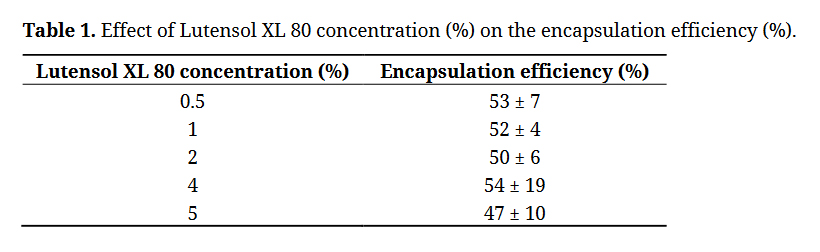 Table 1. Effect of Lutensol XL 80 concentration (%) on the encapsulation efficiency (%).
Poloxamer 188 is considered as valuable alternatives for PVA [9]. Wolf et al. [13] could prepare nanoparticles smaller than 0.4 m in average diameter, and we could also produce particles with similar size using 2% w/v poloxamer 188 with an entrapment efficiency of 70% previously [4]. However, the particles prepared by poloxamer 188 agglomerated after centrifugation similarly to the case shown for PVA emulsifier [4]. In the present work, to increase the relatively low encapsulation efficiency, 2% w/v Lutensol XL 80 was co-applied with 2% w/v poloxamer 188. The resulted entrapment efficiency of the as prepared particles was significantly higher, 77.2 ± 9.3% than that found with each of the emulsifiers.
Size, MorphologyThe amount of stabilizer used has a substantial effect also on the size of the nanoparticles. Most importantly, if the concentration of the stabilizer is too low, aggregation of the polymer droplets will occur and this results in big particles. Too much emulsifier can diminish the encapsulation efficiency. However, when the stabilizer concentration is between these “limits”, adjusting the concentration can be a means of controlling nanoparticle size. In most of the studies investigating protein microencapsulation by double emulsion method, PVA is used as surface active agent [1,4–6,9,14]. Beside its good emulsifier properties, during the washing process, it was found that the synthesized particles agglomerated, and could not be redispersed easily. The aggregation should be avoided during the process, because it may detrimentally influence the application possibility of the particles. Poloxamer is another often used emulsifier in the formation of nano- and micro-particles. Poloxamer is suggested to substitute the PVA in order to avoid agglomeration after lyophilisation [13]. Further, poloxamer have been shown to reduce capture by macrophages and increase the time for systemic circulation [15]. However, in our previous investigations [4] poloxamer was as efficient as PVA in microencapsulation only if using very high concentration of it, moreover, low concentration of poloxamer would be necessary for higher negative zeta potential [16], that is, higher stability. In fact, we realized that poloxamer could not completely inhibit the adhering of the particles after centrifugation. On the contrary, Lutensol XL 80 was suitable for preparing particles with diameter around 1 μm, which did not show aggregation at all. Nevertheless, it must be emphasized this emulsifier was not as efficient in encapsulation efficiency and size decreasing (Tables 1 and 2) as PVA or poloxamer [13]. With this latter two surface active agents, submicron sized particles could be formed easily [4]. Particles with a mean diameter of approximately 1 μm could be formulated by using minimum 2% w/v of Lutensol XL 80 in the external water phase, and this average size could not be substantially decreased by increasing the emulsifier concentration (Table 2, Figure 1).
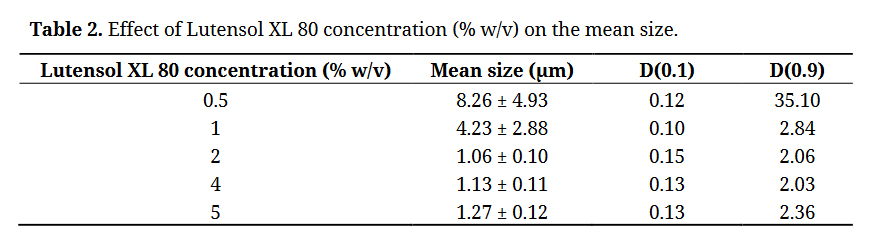 Table 2. Effect of Lutensol XL 80 concentration (% w/v) on the mean size.
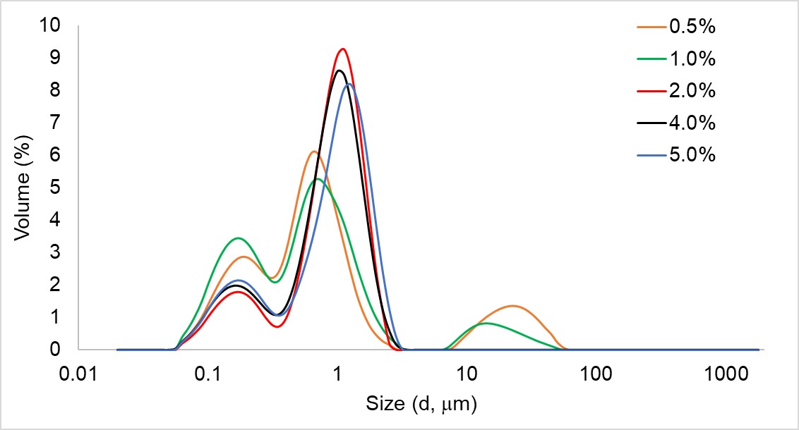 Figure 1. Size distribution of PLGA-HSA composite particles as a function of varying concentration of Lutensol XL80 emulsifier.
As shown above the combination of Lutensol XL 80 with poloxamer 188 emulsifier improved the encapsulation efficiency significantly, thus, its effect on the particle size was also examined. However, the co-use of 2% w/v Lutensol XL 80 and 2% w/v poloxamer provided slightly bigger mean size (1.50 ± 0.14 μm) compared to that of particles formed by the sole application of 2% w/v Lutensol XL 80 (1.06 ± 0.10 μm), the size distribution showed a bit higher amount of the smaller fraction of the particles, and significantly higher size in the bigger particles.
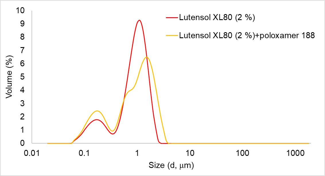 Figure 2. Size distribution of PLGA-HSA composite particles prepared by Lutensol XL80 emulsifier and by its combination with poloxamer 188.
The substantially bigger size of most of the particles is more spectacular in the SEM images (Figure 3). It must be noted the particles were easily redispersable only if the concentration of Lutensol XL 80 was not exceeded by the poloxamer 188 concentration.
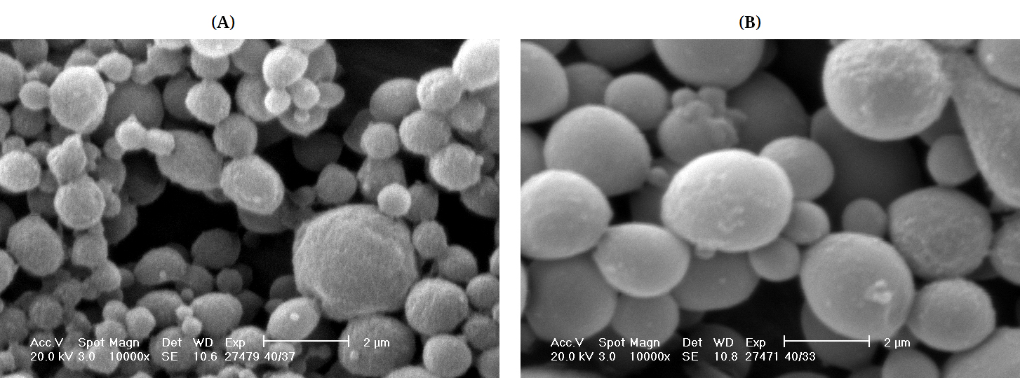 Figure 3. SEM images of particles prepared by Lutensol XL 80 (A) and the combination of Lutensol XL 80 and poloxamer 188 (B).
The shape of the prepared particles was mostly spherical with the Lutensol XL 80 or the emulsifier mixture. However, the sole application of Lutensol XL 80 provided more porous surface to the particles (Figure 3A), while that of the microspheres prepared by the combination of the emulsifiers possessed smoother surface.
Protein Release KineticsHSA release of the model drug-loaded composites was studied for 5 days (Figure 4). The initial burst of HSA from particles formed using Lutensol XL 80 and with its combination with poloxamer 188 was found to be 18.3 ± 4.7% and 13.7 ± 2.3%, respectively, that can be the result of poorly entrapped drug, or drug adsorbed on the surface of the particles [17]. Till the end of the release test, the particles prepared by exclusively Lutensol XL 80 emulsifier released significantly higher amount of protein (30.5 ± 3.4%) compared to that of the composites prepared by the emulsifiers combination (17.8 ± 0.2%). The faster delivery of the particles by Lutensol XL 80 might be explained by their more porous surface observed in the SEM images (Figure 3). An alternative explanation may be the difference in the specific surface area between the two batches. To the smaller size achieved using Lutensol XL 80 belongs larger area/mass and hence shorter diffusion distances and larger fraction of surface bound HSA.
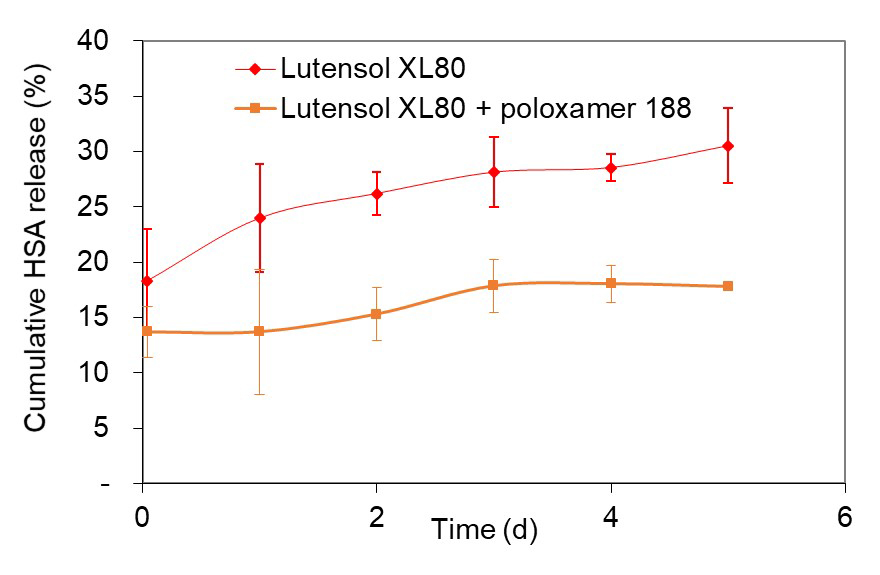 Figure 4. HSA release of particles prepared by Lutensol XL 80 (red) and the combination of Lutensol XL 80 and poloxamer 188 (orange).
PLGA-HSA composite particles were prepared by a novel readily biodegradable emulsifier, Lutensol XL 80 in order to improve controlled drug delivery systems. Particles with mean size of around 1 μm could be formulated. Lutensol XL 80 was found to be a suitable emulsifier in the double emulsion method, although its emulsifying ability was weaker than that of PVA or poloxamer. The relatively low encapsulation efficiency could be increased by combining the conventional emulsifiers with the new surfactant. The main benefit of the Lutensol XL 80 is the improved redispersability of the formed particles after centrifugation compared to the conventional emulsifiers, such as PVA or poloxamer 188. This property could be saved when the Lutensol XL 80 was used in combination with poloxamer 188 in suitable ratios. The release studies implied slow delivery of protein in all of the investigations. Since this new surfactant has not been investigated as material in drug delivery, its potential toxicological effects are not known, thus, further studies are required to ascertain its toxicity.
The author declares that there is no conflict of interest.
The authors greatly acknowledge the financial support given by the BIONANO_GINOP-2.3.2-15-2016-00017 project of the European Structural and Investments Funds and the Hungarian Government.
1.
2.
3.
4.
5.
6.
7.
8.
9.
10.
11.
12.
13.
14.
15.
16.
17.
Feczkó T. Preparation of Model Protein-Loaded PLGA Microparticles Using Lutensol XL 80 Emulsifier. Pharm Front. 2020;2:e200001. https://doi.org/10.20900/pf20200001

Copyright © 2020 Hapres Co., Ltd. Privacy Policy | Terms and Conditions